Review
. 2022 Aug 23;7(1):296.
doi: 10.1038/s41392-022-01132-6.

Classical epithelial-mesenchymal transition (EMT) and alternative cell death process-driven blebbishield metastatic-witch (BMW) pathways to cancer metastasis

Affiliations
Review

Classical epithelial-mesenchymal transition (EMT) and alternative cell death process-driven blebbishield metastatic-witch (BMW) pathways to cancer metastasis

Goodwin G Jinesh et al. Signal Transduct Target Ther. .

Abstract

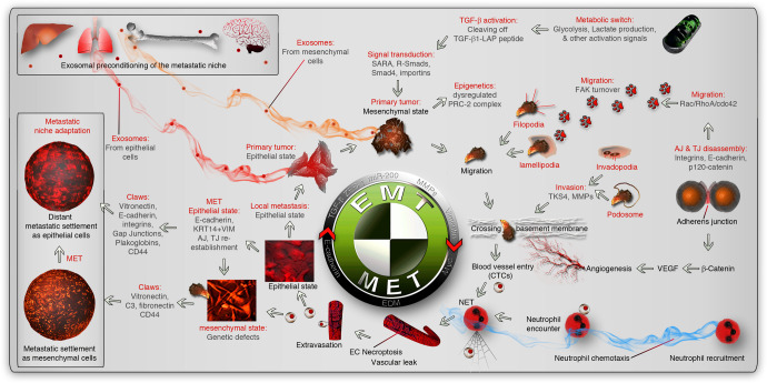
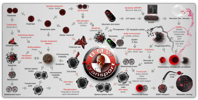
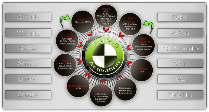
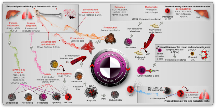
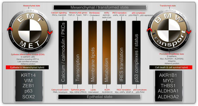
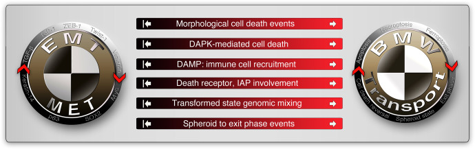
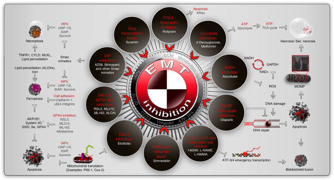
Metastasis is a pivotal event that accelerates the prognosis of cancer patients towards mortality. Therapies that aim to induce cell death in metastatic cells require a more detailed understanding of the metastasis for better mitigation. Towards this goal, we discuss the details of two distinct but overlapping pathways of metastasis: a classical reversible epithelial-to-mesenchymal transition (hybrid-EMT)-driven transport pathway and an alternative cell death process-driven blebbishield metastatic-witch (BMW) transport pathway involving reversible cell death process. The knowledge about the EMT and BMW pathways is important for the therapy of metastatic cancers as these pathways confer drug resistance coupled to immune evasion/suppression. We initially discuss the EMT pathway and compare it with the BMW pathway in the contexts of coordinated oncogenic, metabolic, immunologic, and cell biological events that drive metastasis. In particular, we discuss how the cell death environment involving apoptosis, ferroptosis, necroptosis, and NETosis in BMW or EMT pathways recruits immune cells, fuses with it, migrates, permeabilizes vasculature, and settles at distant sites to establish metastasis. Finally, we discuss the therapeutic targets that are common to both EMT and BMW pathways.


Conflict of interest statement

The authors declare no competing interests.

Figures

Fig. 1
Fig. 1
Classical EMT pathway for metastasis. The metastatic events start with the primary tumor producing exosome-mediated or alternative preconditioning of the metastatic path and niche (indicated by red/orange smoky trail). The metastatic environment is indicated by inset boxes. Altered glycolysis in primary tumor cells or other ECM remodeling triggers TGF-β activation by processing the latency-associate peptide (LAP) to regulate transcription by activation of receptor-regulated Smads-2/3 (R-Smads) through Smad anchor for receptor activation (SARA). Resultant EMT-inducing transcription factors induce a mesenchymal transition from epithelial phenotype while suppressing apoptosis through dysregulated polycomb repressor complex-2 (PRC2). Mesenchymal phenotype is associated with adherens junction (AJ) and tight junction (TJ) disassembly to promote migration (through FAK turnover, lamellipodia, and filopodia) and invasion (through invadopodia and podosomes to cross basement membranes) phenotypes to reach the circulation or through promoting local angiogenesis by VEGF. Inside the blood vessel, neutrophils are attracted by chemotaxis (blue smoky trail), which promotes neutrophil extracellular trap (NET) and necroptosis to facilitate vascular exit. This exposes the circulating tumor cells (CTCs) to a new microenvironment which may or may not promote the reversal of EMT (MET) through the promotion of the epithelial differentiation module (EDM). The extravasated cells use different adhesion cues (claws) to settle and adapt to the new metastatic niche. The reversibility of EMT may support another cycle of EMT and MET
Fig. 2
Fig. 2
Alternative cell death process-dependent BMW pathway for metastasis. The metastatic events start with the initiation of the reversible cell death process. Upon initiation of apoptosis, an acidic environment, and enhanced endocytosis initiate the cell fusion behavior in apoptotic cells. ARAR1 and K-Ras drive the protection of anti-apoptotic mRNAs (IAPs) and IRES translation. Membrane fusion initiates the reassembly of apoptotic blebs to form blebbishields. Blebbishield-blebbishield fusion results in the transformed spheroid state with multiple copies of genomes inside. The nuclear membrane disappears during this stage. Depending on mitochondrial dysfunction and ATP scarcity, the cells can enter an irreversible secondary necrotic state. In parallel, DAMPs (indicated by black or maroon smoky trails from apoptotic and secondary necrotic cells, respectively) attract immune cells to the scene promoting an immune cell-cancer cell hybrid (analogous to a transformed state). The hybrids acquire migratory capacity and use homing signals of immune cells to reach target organs (BMW transport) and use immune cell identity to evade the immune system to establish metastasis. During the transformed spheroid state, p53 is suppressed from expression to suppress apoptosis. The transformed spheres then reorganize their fusogenic lipid membranes (retracted inside from the surface) and reform the nuclear membrane and release individual polarized cancer cells outside the spheroid (Exit phase). The cells released from the transformed state can be polyploid, aneuploid, or diploid to augment genomic instability and heterogeneity. The exited cells may undergo further round of apoptosis due to reactivation of genomic checkpoints, undergo cell division, or undergo cell cycle arrest to augment genomic instability. DAMP damage-associated molecular patterns, MOMP mitochondrial outer membrane permeabilization, BMW transport blebbishield-mediated metastatic-witch transport, PS phosphatidyl serine, CIN chromosomal instability, HSPs heat-shock proteins
Fig. 3
Fig. 3
Activators and signaling components of the TGF-β signaling pathway. A non-exhaustive list of activators of TGF-β signaling (shown in dark brown circles) where one activator may lead to the convergence with the other (green arrows). The downstream signaling receptors and signal transducers (left panel) and important downstream co-operative molecules regulating epithelial-mesenchymal transition (right panel) are listed as side panels. SARA Smad anchor for receptor activation, TAK1 TGF-β activated kinase-1, MAPKs mitogen-activated protein kinases, PRC2 polycomb-repressive complex-2, AP-1 activator protein-1 (Jun/Fos/Fra), EMT-TFs epithelial-to-mesenchymal transition regulatory transcription factors, MCTs monocarboxylate transporters, ECM extracellular matrix, EPO erythropoietin, AJ adherens junction
Fig. 4
Fig. 4
Preconditioning of the metastatic niche. The epithelial and mesenchymal cells precondition the metastatic niche (marked by inset boxes) by cytokine or chemokine secretion or primarily through exosomes (red and orange smoky trails, respectively. The liver, lymph node, and lung metastatic niche were shown as examples (inset boxes). Microbial preconditioning of the liver niche is shown as an example (pathogenic vir-F containing E. coli), and exosomal and immunological preconditioning is also shown in the liver niche. In the lymph node niche, tumor-educated immune cells (B-cells exposed to HSPA4 from tumors) or VEGF-C induced exhaust of cytotoxic T- cells (CTLs) or lymph-protected tumor cells serve as preconditioning mediators (non-exhaustive list of mechanisms). In the lungs, polarized immune cells (into pro-tumor) precondition the niche by cytokines and lipocalin-2 (LCN2). In the cell death context, DAMPs or iDAMPs induce cytotoxic immune cell exhaustion by AICD or immunosuppression, respectively (magenta and green smoke trails, respectively). AICD activation-induced cell death, EC necroptosis endothelial cell necroptosis, MDSCs myeloid-derived suppressor cells, APCs antigen-presenting cells, PGE2 prostaglandin-E2, TSP1 thrombospondin-1, TLR2 Toll-like receptor-2, IAPs inhibitor of apoptotic proteins, NET neutrophil extracellular trap
Fig. 5
Fig. 5
Similarities between EMT and BMW pathways of metastasis. The non-exhaustive common signatures associated with metastasis in the context of EMT and BMW pathways are represented. The mesenchymal state of the EMT pathway is analogous to the transformed state of the BMW pathway and are executed by similar biological functions but with different tools (Top side of the figure). Likewise, the reversible epithelial state of the EMT pathway is analogous to the exit phase of blebbishield emergency program (BMW pathway) and are executed by similar biological functions but with different tools (Bottom side of the figure). The main biological functions shared by EMT and BMW pathways are represented by vertical bars. The key molecules involved in reversible/hybrid-EMT is shown in the black box (far left) and the ones involved in reversible or cell survival and death hybrid is shown in the black box (far right). PKCs protein kinase-C, TSP1 thrombospondin-1, c-IAPs cellular inhibitors of apoptotic proteins, AKRs aldo-ketoreductases, VEGF-A vascular endothelial growth factor-A, PRMT1 protein arginine methyltransferase-1, ALDH aldehyde dehydrogenases, PRC2 polycomb-repressive complex
Fig. 6
Fig. 6
Differences between EMT and BMW pathways of metastasis. The non-exhaustive signatures that discriminate EMT and BMW pathways are illustrated. The white arrows on the red side of the horizontal bars indicate that these events are involved in the BMW pathway of metastasis. The white inhibited arrows on the black side of horizontal bars indicate, that these events are less or not at all involved in the EMT pathway of metastasis. Note that these events may happen in a non-metastatic context in both pathways. DAPK death-activated protein kinase, IAP inhibitor of apoptotic proteins (c-IAPs-1/2, XIAP), DAMP damage-associated molecular patterns
Fig. 7
Fig. 7
Therapeutic vulnerable points which are common to EMT and BMW pathways of metastasis. The common repurposed drugs (white fonts) that aim to inhibit EMT in the contexts of various biological processes (red font) are indicated within brown rings. The EMT inhibitory drug connection to various cell death pathways are indicated outside the brown rings. Note that the IAPs are involved in the reversibility of various cell death forms. Some pathways, such as NAD+ inhibition and PARP1 inhibition, are highly connected through DNA-damage response (DDR). See text for more details and caution statements. IAPs inhibitor of apoptotic proteins, TCA cycle tricarboxylic cycle, PDE4 phosphodiesterase-4, GPX4 glutathione peroxidase-4, iNOS inducible nitric oxide synthase, NAD nicotinamide adenine dinucleotide, CYLD cylindromatosis, GSH glutathione, MLKL mixed lineage kinase domain-like protein, PARP poly (ADP-ribose) polymerase, ATF activating transcription factor; ATP, adenosine triphosphate, GAPDH glyceraldehyde 3-phosphate dehydrogenase, ROS reactive oxygen species

Similar articles

Cited by

References

    1. Lambert AW, Pattabiraman DR, Weinberg RA. Emerging biological principles of metastasis. Cell. 2017;168:670–691. doi: 10.1016/j.cell.2016.11.037. - DOI - PMC - PubMed
    1. Schwartz RS, Erban JK. Timing of metastasis in breast cancer. N. Engl. J. Med. 2017;376:2486–2488. doi: 10.1056/NEJMcibr1701388. - DOI - PubMed
    1. Mehlen P, Puisieux A. Metastasis: a question of life or death. Nat. Rev. Cancer. 2006;6:449–458. doi: 10.1038/nrc1886. - DOI - PubMed
    1. Steeg PS. Targeting metastasis. Nat. Rev. Cancer. 2016;16:201–218. doi: 10.1038/nrc.2016.25. - DOI - PMC - PubMed
    1. Labelle M, Hynes RO. The initial hours of metastasis: the importance of cooperative host-tumor cell interactions during hematogenous dissemination. Cancer Disco. 2012;2:1091–1099. doi: 10.1158/2159-8290.CD-12-0329. - DOI - PMC - PubMed

Publication types