. 2022 Aug 23;13(1):4937.
doi: 10.1038/s41467-022-32710-5.

De novo protein design of photochemical reaction centers

Affiliations

De novo protein design of photochemical reaction centers

Nathan M Ennist et al. Nat Commun. .

Abstract

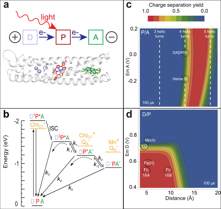
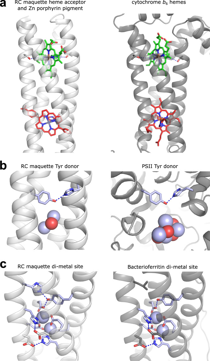
Natural photosynthetic protein complexes capture sunlight to power the energetic catalysis that supports life on Earth. Yet these natural protein structures carry an evolutionary legacy of complexity and fragility that encumbers protein reengineering efforts and obfuscates the underlying design rules for light-driven charge separation. De novo development of a simplified photosynthetic reaction center protein can clarify practical engineering principles needed to build new enzymes for efficient solar-to-fuel energy conversion. Here, we report the rational design, X-ray crystal structure, and electron transfer activity of a multi-cofactor protein that incorporates essential elements of photosynthetic reaction centers. This highly stable, modular artificial protein framework can be reconstituted in vitro with interchangeable redox centers for nanometer-scale photochemical charge separation. Transient absorption spectroscopy demonstrates Photosystem II-like tyrosine and metal cluster oxidation, and we measure charge separation lifetimes exceeding 100 ms, ideal for light-activated catalysis. This de novo-designed reaction center builds upon engineering guidelines established for charge separation in earlier synthetic photochemical triads and modified natural proteins, and it shows how synthetic biology may lead to a new generation of genetically encoded, light-powered catalysts for solar fuel production.


Conflict of interest statement

The authors declare no competing interests.

Figures

Fig. 1
Fig. 1. Design of natural and artificial photosynthetic reaction centers.
a The light-activated donor-pigment-acceptor (DPA) electron transfer triad core of photosynthesis and X-ray crystal structure at 2.0 Å resolution of the RC maquette with a metal ion/tyrosine donor, Zn porphyrin pigment, and heme B acceptor (PDB ID: 5VJS). b Kinetic schemes of a light-activated system show energies plotted relative to ground state before light activation. An RC maquette DPA triad is compared with representative PSII charge separated states (orange). c Triad contour plots of expected relative charge separated D+PA yield after 100 µs for a range of P-to-A edge-to-edge distances vs. acceptor Em (using Em of 0.91 V for P/P+ and Em of 0.72 V for tyrosinate donor). Dashed lines show cofactor anchoring residues adjusted in increments of ~1 helical turn (~5.2 Å). Fe porphyrins heme B and DADPIX are shown as acceptor alternatives. d Corresponding triad contour plot for D-to-P distances and driving forces; acceptor is heme B; tyrosinate, Mn(II), Fe(II) and cysteine-coupled ferrocene (Fc164 and Fc168) shown as possible donors.
Fig. 2
Fig. 2. Structural similarities between cofactor binding sites of designed RC maquette and natural proteins are significant despite lack of sequence identity.
RC maquette crystal structures are shown in white with heme electron acceptor and ligating His residues shown in green, ZnP pigment and His in red, and electron donating Tyr, metal ion spheres and first shell ligands in blue. Natural protein structures are shown in gray with cofactors colored as in RC maquette. Metal-bridging oxygens are shown as red spheres. Blue dotted lines represent hydrogen bonds. a RC maquette (PDB ID: 5VJS) and cytochrome b6f (PDB ID: 6RQF). b RC maquette-L71H mutant with Cd(II) (PDB ID: 5VJU) and PSII (PDB ID: 6DHE). c RC maquette-L71H mutant with Cd(II) (PDB ID: 5VJU) and bacterioferritin with Cd(II) (PDB ID: 4CVS).
Fig. 3
Fig. 3. Transient spectroscopy of RC maquette reveals the dynamics of light-activated charge separation and recombination.
a Difference spectra of the Tyr-ZnP-heme triad in L31D/L71H mutant at pH 9.5 are shown for delay times from 1 μs to 3 s after the laser flash at log time intervals shown in b. b The first and second principal time varying SVD components of the difference spectra (circles and squares, respectively) fit to a simple kinetic model connecting DP*A, DP+A, D+PA, and DPA states with single exponential first order reactions (solid lines): ZnP monad (red), ZnP-Heme dyad (green), Tyr-ZnP-heme triad (blue), (see also Supplementary Figs. 4 and 6). c Fitted log rates for the Tyr-ZnP-heme triad. d Fitted DP*A and D+PA difference spectra for the Tyr-ZnP-heme triad dominated by ZnP bleach (red) and heme redox (blue) difference spectra, respectively, compared to a scaled heme redox spectrum acquired for a 20 µM RC maquette-heme sample reduced with dithionite in the dark (black dashed line). e Fitted time varying SVD components for the ferrocene-ZnP-Heme triad (cyan) and iron-Tyr-ZnP-heme tetrad (orange) show charge separation lifetimes up to 300 ms. Source data are provided as a Source Data file.

References

    1. Moser CC, Keske JM, Warncke K, Farid RS, Dutton PL. Nature of biological electron transfer. Nature. 1992;355:796–802. doi: 10.1038/355796a0. - DOI - PubMed
    1. Moser CC, Page CC, Leslie Dutton P. Tunneling in PSII. Photochem. Photobio. Sci. 2005;4:933–939. doi: 10.1039/b507352a. - DOI - PubMed
    1. Moser CC, Anderson JL, Dutton PL. Guidelines for tunneling in enzymes. Biochim. Biophys. Acta. 2010;1797:1573–1586. doi: 10.1016/j.bbabio.2010.04.441. - DOI - PMC - PubMed
    1. Barber J, Tran PD. From natural to artificial photosynthesis. J. R. Soc. Interface. 2013;10:20120984. doi: 10.1098/rsif.2012.0984. - DOI - PMC - PubMed
    1. Vinyard DJ, Brudvig GW. Progress toward a molecular mechanism of water oxidation in photosystem II. Annu Rev. Phys. Chem. 2017;68:101–116. doi: 10.1146/annurev-physchem-052516-044820. - DOI - PubMed

Publication types

Substances