. 2022 Aug 23;12(1):14345.
doi: 10.1038/s41598-022-18323-4.

Mental imagery of whole-body motion along the sagittal-anteroposterior axis

Affiliations

Mental imagery of whole-body motion along the sagittal-anteroposterior axis

K Patel et al. Sci Rep. .

Abstract

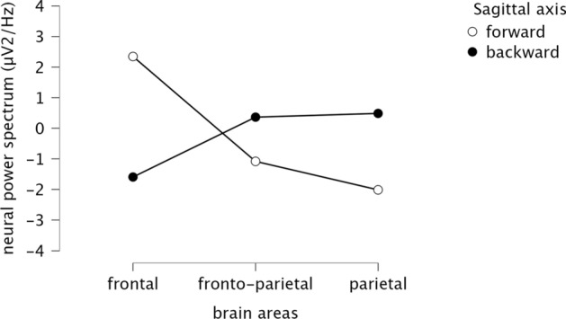
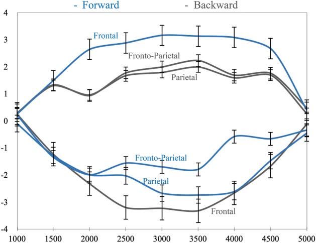
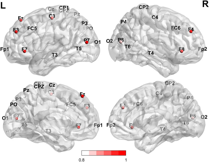
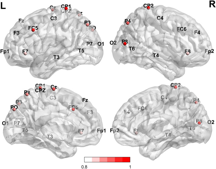
Whole-body motor imagery is conceptualised as a mental symbolisation directly and indirectly associated with neural oscillations similar to whole-body motor execution. Motor and somatosensory activity, including vestibular activity, is a typical corticocortical substrate of body motion. Yet, it is not clear how this neural substrate is organised when participants are instructed to imagine moving their body forward or backward along the sagittal-anteroposterior axis. It is the aim of the current study to identify the fingerprint of the neural substrate by recording the cortical activity of 39 participants via a 32 electroencephalography (EEG) device. The participants were instructed to imagine moving their body forward or backward from a first-person perspective. Principal Component Analysis (i.e. PCA) applied to the neural activity of whole-body motor imagery revealed neural interconnections mirroring between forward and backward conditions: beta pre-motor and motor oscillations in the left and right hemisphere overshadowed beta parietal oscillations in forward condition, and beta parietal oscillations in the left and right hemisphere overshadowed beta pre-motor and motor oscillations in backward condition. Although functional significance needs to be discerned, beta pre-motor, motor and somatosensory oscillations might represent specific settings within the corticocortical network and provide meaningful information regarding the neural dynamics of continuous whole-body motion. It was concluded that the evoked multimodal fronto-parietal neural activity would correspond to the neural activity that could be expected if the participants were physically enacting movement of the whole-body in sagittal-anteroposterior plane as they would in their everyday environment.


Conflict of interest statement

The authors declare no competing interests.

Figures

Figure 1
Figure 1
Schematic presentation of the mental preparation in two steps. (a) The experimenter moves the chair forward and backward in front of each participant first, and ask the participant to do the same immediately after; (b) the experimenter sits in the chair and moves forward and backward while sitting, and asked each participant to repeat the same.
Figure 2
Figure 2
Timing presentation of the experimental paradigm of the whole body motor imagery. The start of each trial was signified by the presence of a short message “Ready”. This corresponds to t = 0. After, a fixation point appeared in the middle of the screen for 2 s, i.e., t = 1, fixation. After these 2 s, a direction-phrase indicating either "go forward" or "go backward" appeared for 2 s on the screen (i.e., t = 2). Once the words disappeared from the screen, each participant, facing the black screen, performed a motor imagery task, one at a time for 5 s, i.e., t = 3. At the end of each motor imagery task, each participant was allowed to take a break "please wait", i.e. t = 4 for 2 s. In total, for 39 participants, there were 1,872 randomised trials. In 1,872 trials of 5 s each, resulted in 9360 s worth of data.
Figure 3
Figure 3
The experimenter instructed the participants to perform the mental imagery of whole-body motion from a first-person perspective: imagine move forward vs backward one at a time. To that end, the experimenter explained the paradigm to the participants step by step (i.e. from t = 0 to t = 4) verbally assisting each participant to respect the chronology and directives of each step (i.e. fix, read, imagine, relax) within the trials (i.e. experimenter-participant training). Each participant was also assisted to perform the task by him/herself as well (i.e. participant training). Participants were equipped with 32-electrode wireless EEG system.
Figure 4
Figure 4
The participants were sitting in the same chair that s/he used for the mental preparation phase, the baseline and the training phase. Along with the training phase, each participant was adjusted in the front of an LCD screen monitor and instructed to imagine his/her own body motion when appropriate. The experimental paradigm was the same as presented in the training phase. All participants performed the mental imagery task in the dark. Participants were equipped with 32-electrode wireless EEG system.
Figure 5
Figure 5
Comparison between forward and backward motor imagery (i.e. sagittal axis) in relationship with the average power spectrum in the three beta-frequency ROIs. X axis represents the selected ROIs (i.e. frontal, fronto-parietal and parietal); Y axis illustrates the average neural power spectrum (μV2/Hz) in beta-frequency (13.5–30 Hz), i.e. power spectrum of the neural activity.
Figure 6
Figure 6
Temporal evolution of beta-frequency (13.5–30 Hz) over 5 s time period across the three ROI’s for forward and backward mental imagery (i.e. sagittal axis). X axis represents the time-course of 5 s in milliseconds based on a time-window of 1000 ms; Y axis illustrates the average power spectrum of the neural activity (μV2/Hz). Blue colour depicts "forward motor imagery"; grey colour illustrates "backward motor imagery".
Figure 7
Figure 7
Loadings derives from PCA (Principal Component Analysis) for forward mental imagery. The PCA revealed two components accounting for 76% of the total variance. There was more beta synchronisation in frontal bilateral areas and less beta synchronisation in fronto-parietal unilateral left areas. Bilateral parietal synchronisation was also observed (Top: Sagittal external section; Bottom: internal sections. L: left hemisphere; R: right hemisphere).
Figure 8
Figure 8
Loadings derives from PCA (Principal Component Analysis) for backward mental imagery. Two components which explained 94% of the total variance were reported. There was more beta synchronisation in parietal and fronto-parietal areas bilaterally and less beta synchronisation in frontal areas bilaterally and unilaterally left areas (Top: Sagittal external section; Bottom: internal sections. L: left hemisphere; R: right hemisphere).

References

    1. Kilteni K, Andersson BJ, Houborg C, Ehrsson HH. Motor imagery involves predicting the sensory consequences of the imagined movement. Nat. Commun. 2018;9:1617. doi: 10.1038/s41467-018-03989-0. - DOI - PMC - PubMed
    1. Jeannerod M. Neural simulation of action: A unifying mechanism for motor cognition. Neuroimage. 2001;14:S103–109. doi: 10.1006/nimg.2001.0832. - DOI - PubMed
    1. Ehrsson HH, Geyer S, Naito E. Imagery of voluntary movement of fingers, toes, and tongue activates corresponding body-part-specific motor representations. J. Neurophysiol. 2003;90:3304–3316. doi: 10.1152/jn.01113.2002. - DOI - PubMed
    1. Giannopulu I, Mizutani H. Neural kinesthetic contribution to motor imagery of body parts: Tongue, hands, and feet. Front. Hum. Neurosci. 2021;15:602723. doi: 10.3389/fnhum.2021.602723. - DOI - PMC - PubMed
    1. Brandt T, Bartenstein P, Janek A, Dieterich M. Reciprocal inhibitory visual-vestibular interaction. Visual motion stimulation deactivates the parieto-insular vestibular cortex. Brain. 1998;121(Pt 9):1749–1758. doi: 10.1093/brain/121.9.1749. - DOI - PubMed