Review
. 2022 Aug 23;20(1):383.
doi: 10.1186/s12951-022-01584-6.

Tumor-derived small extracellular vesicles: potential roles and mechanism in glioma

Affiliations
Review

Tumor-derived small extracellular vesicles: potential roles and mechanism in glioma

Xu Guo et al. J Nanobiotechnology. .

Abstract

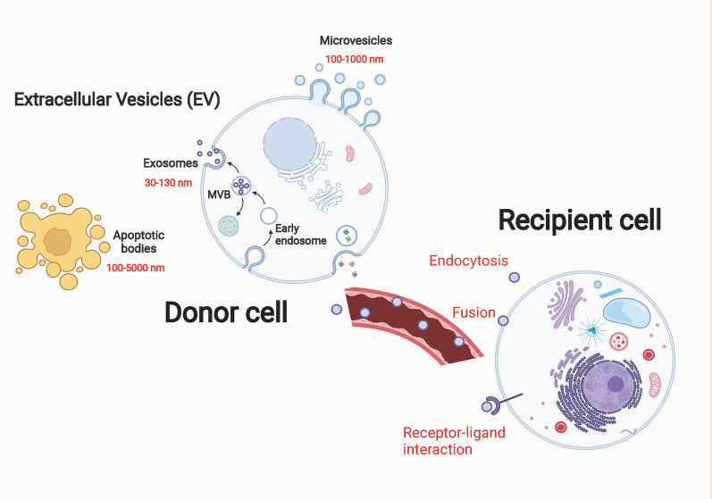
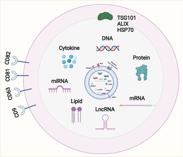
Small extracellular vesicles (SEVs) are extracellular vesicles containing DNA, RNA, and proteins and are involved in intercellular communication and function, playing an essential role in the growth and metastasis of tumors. SEVs are present in various body fluids and can be isolated and extracted from blood, urine, and cerebrospinal fluid. Under both physiological and pathological conditions, SEVs can be released by some cells, such as immune, stem, and tumor cells, in a cytosolic manner. SEVs secreted by tumor cells are called tumor-derived exosomes (TEXs) because of their origin in the corresponding parent cells. Glioma is the most common intracranial tumor, accounting for approximately half of the primary intracranial tumors, and is characterized by insidious onset, high morbidity, and high mortality rate. Complete removal of tumor tissues by surgery is difficult. Chemotherapy can improve the survival quality of patients to a certain extent; however, gliomas are prone to chemoresistance, which seriously affects the prognosis of patients. In recent years, TEXs have played a vital role in the occurrence, development, associated immune response, chemotherapy resistance, radiation therapy resistance, and metastasis of glioma. This article reviews the role of TEXs in glioma progression, drug resistance, and clinical diagnosis.

Keywords: Glioma; Immunotherapies; Malignant progression; Tumor-derived SEVs.


Conflict of interest statement

The authors declare no competing interests.

Figures

Fig. 1
Fig. 1
The main process of exosome biogenesis and release. Most cells in human body can release double-layer membrane-bound nanovesicles into the extracellular space. These membrane-derived vesicles can be divided into three types according to their size: small extracellular vesicles, microvesicles, and apoptotic bodies. Small extracellular vesicles originate from intracellular multivesicular bodies. Multivesicular bodies are vesicles with endocytosis. After fusion with the plasma membrane, a part is degraded by lysosomes, and the other part is released outside the cell to form small extracellular vesicles. Three main ways of information transmission exist between small extracellular vesicles and target cells. Exosome membrane proteins interact with target cell membrane proteins to activate intracellular signaling pathways. Small extracellular vesicles can transfer their own genetic material. The exosome membrane can be directly fused with the target cell membrane, and the genetic information carried in the exosome can be directly transferred to the recipient cell
Fig. 2
Fig. 2
The contents of small extracellular vesicles. Almost all types of cells can secrete small extracellular vesicles, and small extracellular vesicles widely contain nucleic acids such as microRNA (miRNA), lncRNA, circRNA, mRNA, proteins, and lipids. Their surface markers mainly include CD63, CD81, CD9, ALG-2 Interacting protein X (Alix), tumor susceptibility gene 101 (TSG101), and heat shock protein 70 (HSP70)
Fig. 3
Fig. 3
The cells involved in the tumor microenvironment. The tumor microenvironment is the growth environment of tumor cells, which is composed of tumor cells, immune cells, interstitial cells, extracellular matrix, and other factors, whose composition is more complex
Fig. 4
Fig. 4
The involving participants mediating the radiation- and chemoresistance of glioma
Fig. 5
Fig. 5
The extracellular matrix in GBM tumorigenesis. The features of extracellular matrix in normal brain are low overall density and stiffness and absence of neurodevelopmental proteins. The features of extracellular matrix in GBM are increased overall density and stiffness, presence of oncofetal protein isoforms, and promotion of invasion and angiogenesis
Fig. 6
Fig. 6
The features of tumor angiogenesis. Tumor angiogenesis is a complex process of interaction between tumor cells and endothelial cells. The main process is as follows: endothelial cells are stimulated by proangiogenic growth factors secreted by tumor cells. After degrading, endothelial cells proliferate under the action of various chemokines, form lumen with pericytes under the action of adhesion molecules, and generate new tumor blood vessels after maturation and stabilization. Tumor angiogenesis is an important basis for tumor growth, invasion, and metastasis. This process is regulated by various angiogenic growth-promoting or inhibitory factors and signaling pathways

References

    1. Yang K, Wu Z, Zhang H, Zhang N, Wu W, Wang Z, Dai Z, Zhang X, Zhang L, Peng Y, Ye W, Zeng W, Liu Z, Cheng Q. Glioma targeted therapy: insight into future of molecular approaches. Mol Cancer. 2022;21:39. doi: 10.1186/s12943-022-01513-z. - DOI - PMC - PubMed
    1. Zhou Q, Xue C, Ke X, Zhou J. Treatment response and prognosis evaluation in high-grade glioma: an imaging review based on MRI. J Magn Reson Imaging. 2022;56:325–340. doi: 10.1002/jmri.28103. - DOI - PubMed
    1. Ho RLY, Ho IAW. Recent advances in glioma therapy: combining vascular normalization and immune checkpoint blockade. Cancers (Basel) 2021;13:3686. doi: 10.3390/cancers13153686. - DOI - PMC - PubMed
    1. Takacs GP, Flores-Toro JA, Harrison JK. Modulation of the chemokine/chemokine receptor axis as a novel approach for glioma therapy. Pharmacol Ther. 2021;222:107790. doi: 10.1016/j.pharmthera.2020.107790. - DOI - PMC - PubMed
    1. Tamai S, Ichinose T, Tsutsui T, Tanaka S, Garaeva F, Sabit H, Nakada M. Tumor microenvironment in glioma invasion. Brain Sci. 2022;12:505. doi: 10.3390/brainsci12040505. - DOI - PMC - PubMed