

Highly Cellular Leiomyoma Mixed With a Focus of Adenomyosis
- PMID: 35999995
- PMCID: PMC9390799
- DOI: 10.7759/cureus.28129
Highly Cellular Leiomyoma Mixed With a Focus of Adenomyosis
Abstract
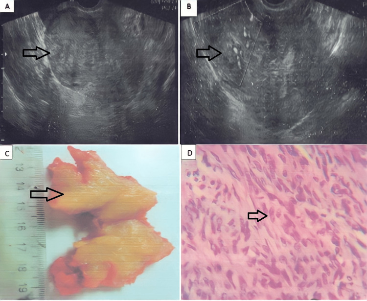
The purpose of this case presentation was to highlight the importance of an adequate evaluation of images when suspicious of atypical leiomyoma and the importance of performing an extemporaneous biopsy during surgery to ensure the lesion is a benign muscular cell tumor. Here, we present a case of a 34-year-old nulliparous woman who presented with a history of infertility and irregular menstrual cycles. A highly vascularized pelvic mass was visualized by Doppler ultrasound and a contrast MRI suggestive of uterine fibroid. Intraoperatively, the lesion was found adjacent to the uterus, with a second lesion deeper into the myometrium. The pathology reported a highly cellular leiomyoma with a focus of adenomyosis. Both lesions were extirpated without complications. The patient recuperated favorably within three months of follow-up. This case shows an example of a variety of the typical histology that can be found in uterine fibroids. Although the management of atypical leiomyomas could vary in different scenarios, conservative treatment is recommended if fertility wishes are present. In all cases, it is mandatory to exclude any possibilities of malignancy, like sarcoma, which would completely change the intraoperative management.
Keywords: adenomyosis; fibroids; high cellularity; leiomyoma; leiomyosarcoma.
Copyright © 2022, Nava et al.
Conflict of interest statement
The authors have declared that no competing interests exist.
Figures
References
-
- Diagnostic algorithm to differentiate benign atypical leiomyomas from malignant uterine sarcomas with diffusion-weighted MRI. Abdel Wahab C, Jannot AS, Bonaffini PA, et al. Radiology. 2020;297:361–371. - PubMed
-
- Review of uterine fibroids: imaging of typical and atypical features, variants, and mimics with emphasis on workup and FIGO classification. Awiwi MO, Badawy M, Shaaban AM, et al. Abdom Radiol (NY) 2022;47:2468–2485. - PubMed
Publication types
LinkOut - more resources
Full Text Sources