. 2022 Oct 26;13(5):e0200422.
doi: 10.1128/mbio.02004-22. Epub 2022 Aug 24.

Mycobacterium tuberculosis Utilizes Host Histamine Receptor H1 to Modulate Reactive Oxygen Species Production and Phagosome Maturation via the p38MAPK-NOX2 Axis

Affiliations

Mycobacterium tuberculosis Utilizes Host Histamine Receptor H1 to Modulate Reactive Oxygen Species Production and Phagosome Maturation via the p38MAPK-NOX2 Axis

Siwei Mo et al. mBio. .

Abstract

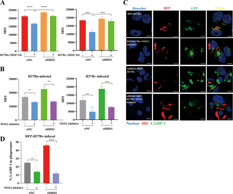
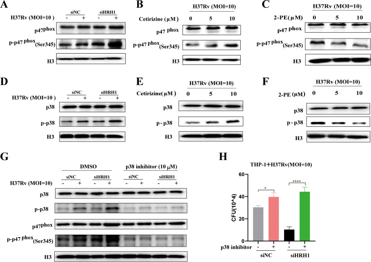
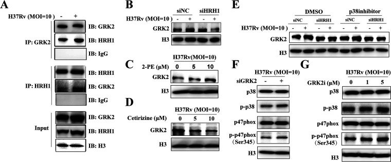
Tuberculosis (TB), which is caused by the single pathogenic bacterium, Mycobacterium tuberculosis, is among the top 10 lethal diseases worldwide. This situation has been exacerbated by the increasing number of cases of multidrug-resistant TB (MDR-TB) and extensively drug-resistant TB (XDR-TB). Histamine is an organic nitrogenous compound that mediates a plethora of cell processes via different receptors. The expression of histamine receptor H1 (HRH1), one of the four histamine receptors identified to date was previously reported to be augmented by M. tuberculosis infection, although the underlying mechanism is unclear. In the present study, we applied confocal microscopy, flow cytometry, and Western blotting to show that HRH1 expression was enhanced in macrophages following mycobacterial infection. Furthermore, by combining techniques of gene knockdown, immunoprecipitation, intracellular bacterial burden analysis, fluorescence labeling, and imaging, we found that M. tuberculosis targeted the host HRH1 to suppress NOX2-mediated cROS production and inhibit phagosome maturation and acidification via the GRK2-p38MAPK signaling pathway. Our findings clarified the underlying mechanism of the M. tuberculosis and host HRH1 interaction and may provide useful information for the development of novel antituberculosis treatments. IMPORTANCE Once engulfed in macrophage phagosomes, M. tuberculosis adopts various strategies to take advantage of the host environment for its intracellular survival. Histamine is an organic nitrogen-containing compound that mediates a plethora of cellular processes via different receptors, but the crosstalk mechanism between M. tuberculosis and HRH1 in macrophages is not clear. Our results revealed that M. tuberculosis infection enhanced HRH1 expression, which in turn restrained macrophage bactericidal activity by modulating the GRK2-p38MAPK signaling pathway, inhibiting NOX2-mediated cROS production and phagosome maturation. Clarification of the underlying mechanism by which M. tuberculosis utilizes host HRH1 to favor its intracellular survival may provide useful information for the development of novel antituberculosis treatments.

Keywords: HRH1; Mycobacterium tuberculosis; NOX2; ROS; p38MAPK.


Conflict of interest statement

The authors declare no conflict of interest.

Figures

FIG 1
FIG 1
Histamine-activated HRH1 signaling enhanced the intracellular survival of M. tuberculosis in macrophages. (A) Colony forming unit (CFU) analysis of intracellular mycobacteria in PMA-differentiated THP-1 macrophages infected with H37Ra or H37Rv (MOI = 10:1) for 6 h, and then treated with 10 μM histamine for another 72 h. (B) Western blotting of protein expression levels of the four histamine receptors in THP-1 macrophages infected with H37Ra or H37Rv (MOI = 10:1) for 24 h. (C) RT-qPCR analysis of THP-1 macrophages infected with H37Ra or H37Rv (MOI = 10:1) for 24 h. (D) Flow cytometric analysis of HRH1 expression in THP-1 macrophages infected with H37Ra or H37Rv (MOI = 10:1); data represent HRH1+ cell counts and HRH1 mean fluorescence intensity (MFI). (E) Representative immunofluorescence confocal microscopy images (total 80 to 100 images, scale bars: 5 μm) of HRH1 expression in THP-1 macrophages infected with GFP labeled H37Ra (MOI = 10:1). All data represent the mean ± SD of at least three experiments. *, P < 0.05; **, P < 0.01; ***, P < 0.001; ****, P < 0.0001; ns, not significant.
FIG 2
FIG 2
Inhibition or knockdown of HRH1 attenuated M. tuberculosis survival in macrophages. (A) Colony forming unit (CFU) analysis of intracellular mycobacteria in PMA-differentiated THP-1 macrophages infected with H37Ra or H37Rv for 6 h, and then treated with 10 μM 2-PE (HRH1 agonist) for 72 h. (B) Colony forming unit (CFU) analysis of intracellular mycobacteria in PMA-differentiated THP-1 macrophages infected with H37Ra or H37Rv (MOI = 10:1) for 6 h before treatment with 10 μM cetirizine (HRH1 antagonist) for 72 h. (C) Western blotting of histamine receptor (HRH1 to 4) protein expression level in THP-1 cells transfected for 48 h with gene-targeted siRNAs. (D) Colony forming unit (CFU) analysis of intracellular mycobacteria in PMA-differentiated THP-1 macrophages transfected for 48 h with gene-targeted siRNAs before infection with H37Ra (MOI = 10:1) for 72 h. (E) Colony forming unit (CFU) analysis of intracellular mycobacteria in PMA-differentiated THP-1 macrophages transfected for 48 h with gene-targeted siRNAs before infection with H37Rv (MOI = 10:1) for 72 h. All data represent the mean ± SD of at least three experiments. *, P < 0.05; **, P < 0.01; ***, P < 0.001; ****, P < 0.0001; ns, not significant.
FIG 3
FIG 3
HRH1 suppressed NOX2-mediated cROS production. (A) PMA-differentiated THP-1 macrophages were transfected with HRH1 or control siRNA for 48 h before infection with H37Ra (MOI = 10:1) for 24 h. Cytoplasmic ROS (cROS) was analyzed by flow cytometry. (B) PMA-differentiated THP-1 macrophages were transfected with HRH1 or control siRNA for 48 h before infection with H37Rv (MOI = 10:1) for 24 h. Cytoplasmic ROS (cROS) was analyzed by flow cytometry. (C) Western blotting of the protein expression level of NOX family members following either infection with H37Ra and H37Rv alone or in combination and with or without HRH1 knockdown. (D) PMA-differentiated THP-1 macrophages were transfected with HRH1 or control siRNA for 48 h before infection with H37Ra (MOI = 10:1) for 24 h. NADPH oxidase activity was analyzed by measuring the absorbance at 450 nm using the EpochTM 2 microplate spectrophotometer. (E) Flow cytometric analysis of cROS expression following infection with H37Ra or H37Rv and in the presence or absence of 25 μM NOX2 inhibitor. (F) Colony forming unit (CFU) analysis of intracellular mycobacteria in THP-1 macrophages with or without 25 μM NOX2 inhibitor. All data represent the mean ± SD of at least three experiments. *, P < 0.05; **, P < 0.01; ***, P < 0.001; ****, P < 0.0001; ns, not significant.
FIG 4
FIG 4
M. tuberculosis infection inhibits phagosome maturation and acidification via the HRH1-NOX2 axis. (A) PMA-differentiated THP-1 macrophages were transfected with HRH1 or control siRNA for 48 h before infection with H37Ra or H37Rv (MOI = 10:1) for another 24 h. Flow cytometric analysis of intracellular phagosome acidification levels. (B) PMA-differentiated THP-1 macrophages were transfected with HRH1 or control siRNA for 48 h before infection with H37Ra or H37Rv (MOI = 10:1) and cultured with or without the NOX2 inhibitor (25 μM) for another 24 h. Flow cytometric analysis of intracellular phagosome acidification levels. (C) Representative immunofluorescence confocal microscopy images (total 80 to 100 images) to illustrate the staining of phagosome maturation in RFP-H37Rv infection macrophages; scale bars: 5 μm. (D) Quantification of phagosome maturation using LAMP-1 as an indicator. All data represent the mean ± SD of at least three experiments. **, P < 0.01; ***, P < 0.001; ****, P < 0.0001; ns, not significant.
FIG 5
FIG 5
HRH1 inhibited p47phox phosphorylation to suppress NOX2 activity via the p38MAPK signaling pathway. (A) Western blotting of the protein levels of p47phox and p-p47phox (Ser345) in PMA-differentiated THP-1 cells transfected with HRH1 or control siRNA for 48 h before infection with H37Rv (MOI = 10:1) for another 24 h. (B) Western blotting of the protein levels of p47phox and p-p47phox (Ser345) in the presence or absence of cetirizine (0 μM, 5 μM, 10 μM). (C) Western blotting of the protein levels of p47phox and p-p47phox (Ser345) in the presence or absence of 2-PE (0 μM, 5 μM, 10 μM). (D) Western blotting of the protein levels of p38MAPK and p-p38MAPK in PMA-differentiated THP-1 cells transfected with HRH1 or control siRNA for 48 h before infection with H37Rv (MOI = 10:1) for another 24 h. (E) Western blotting of the protein levels of p38MAPK and p-p38MAPK in PMA-differentiated THP-1 cells transfected with HRH1 or control siRNA for 48 h and cultured in the presence or absence of cetirizine (0 μM, 5 μM, 10 μM). (F) Western blotting of the protein levels of p38MAPK and p-p38MAPK in the presence or absence of 2-PE (0 μM, 5 μM, 10 μM). (G-H) PMA-differentiated THP-1 cells were transfected with HRH1 or control siRNA for 48 h before infection with H37Rv (MOI = 10:1) for another 24 h in the presence or absence of 10 μM p-p38MAPK inhibitor. (G) Western blotting of the protein levels of p38MAPK, p-p38MAPK, p47phox, and p-p47phox (Ser345). (H) Colony forming unit (CFU) analysis of intracellular bacteria. All data represent the mean ± SD of at least three experiments. *, P < 0.05; ****, P < 0.0001.
FIG 6
FIG 6
HRH1 interacts with GRK2 to negatively regulate the p38MAPK signaling pathway. (A) PMA-differentiated THP-1 cells were infected with H37Rv (MOI = 10:1) for 24 h and immunoprecipitated (IP) with anti-GRK2 or anti-HRH1 antibodies. Immunoprecipitates were then immunoblotted (IB) with anti-GRK2 or anti-HRH1 antibodies, accordingly. (B) Western blotting of GRK2 protein levels in PMA-differentiated THP-1 cells transfected with HRH1 siRNA for 48 h and infected with H37Rv (MOI = 10:1) for 24 h. (C) Western blotting of GRK2 protein levels in the presence or absence of 2-PE (0, 5 μM, 10 μM) with H37Rv (MOI = 10:1) infection for 24 h. (D) Western blotting of GRK2 protein levels in the presence or absence of cetirizine (0, 5 μM, 10 μM) with H37Rv (MOI = 10:1) infection for 24 h. (E) Western blotting of GRK2 protein levels in PMA-differentiated THP-1 cells transfected with HRH1 siRNA for 48 h and infected with H37Rv (MOI = 10:1) for 24 h in the presence or absence of 10 μM p-p38MAPK inhibitor. (F) Western blotting of the protein levels of p47phox/p-p47phox (Ser345), p38MAPK, and p-p38MAPK in PMA-differentiated THP-1 cells transfected with GRK2 siRNA for 48 h and infected with H37Rv (MOI = 10:1) for 24 h. (G) Western blotting of the protein levels of p47phox/p-p47phox (Ser345), p38MAPK, and p-p38MAPK in the presence or absence of GRK2i (inhibitor, 0, 1, or 5 μM) with H37Rv (MOI = 10:1) infection for 24 h.
FIG 7
FIG 7
Mycobacterium tuberculosis utilized host HRH1 to modulate reactive oxygen species production and phagosome maturation via the GRK2-p38MAPK-NOX2 axis, a summary model. Uninfected macrophages displayed normal regulation of HRH1 and p38MAPK pathway (left). After mycobacteria infection and endocytosis by macrophages (middle panel), HRH1 expression was enhanced and interacted with GRK2, which then inhibited the p38MAPK signaling pathway. The inhibited p38MAPK signaling further decreased the level of phosphorylated oxidase subunit p47phox (Ser345), leading to the inability of the oxidase subunit p47phox (Ser345) to bind NOX2 (gp91phox) on the phagosome membrane, and thus inhibited the NOX2 (gp91phox) oxidase complex formation. This reduced cROS production, which further prevented phagosome maturation and acidification. This process provided a beneficial environment that favored the intracellular survival of M. tuberculosis in macrophages. However, HRH1 inhibitor treatment blocked the inhibitory effect of GRK on p38MAPK signaling in M. tuberculosis-infected macrophages. As a result, NOX2 could function normally to promote phagosome maturation and acidification, which was detrimental to the survival of M. tuberculosis in macrophages (right).

Similar articles

Cited by

References

    1. Shah M, Dorman SE. 2021. Latent tuberculosis infection. N Engl J Med 385:2271–2280. doi:10.1056/NEJMcp2108501. - DOI - PubMed
    1. Glickman MS, Jacobs WR, Jr. 2001. Microbial pathogenesis of Mycobacterium tuberculosis: dawn of a discipline. Cell 104:477–485. doi:10.1016/S0092-8674(01)00236-7. - DOI - PubMed
    1. Jeremiah C, Petersen E, Nantanda R, Mungai BN, Migliori GB, Amanullah F, Lungu P, Ntoumi F, Kumarasamy N, Maeurer M, Zumla A. 2022. The WHO Global Tuberculosis 2021 Report - not so good news and turning the tide back to end TB. Int J Infect Dis S1201-9712:149-7. - PMC - PubMed
    1. Brites D, Gagneux S. 2015. Co-evolution of Mycobacterium tuberculosis and Homo sapiens. Immunol Rev 264:6–24. doi:10.1111/imr.12264. - DOI - PMC - PubMed
    1. Ernst JD. 2012. The immunological life cycle of tuberculosis. Nat Rev Immunol 12:581–591. doi:10.1038/nri3259. - DOI - PubMed

Publication types

MeSH terms