

Genome Editing of Veterinary Relevant Mycoplasmas Using a CRISPR-Cas Base Editor System
- PMID: 36000854
- PMCID: PMC9469718
- DOI: 10.1128/aem.00996-22
Genome Editing of Veterinary Relevant Mycoplasmas Using a CRISPR-Cas Base Editor System
Abstract
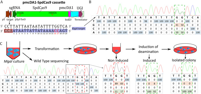
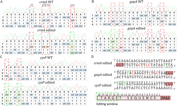
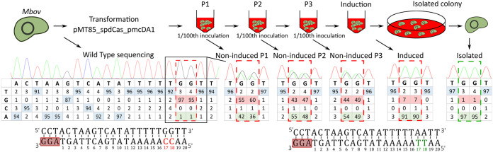
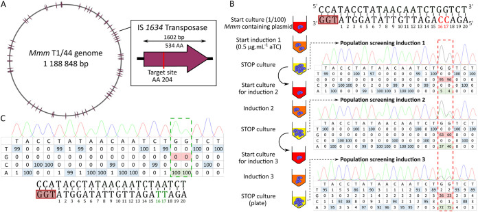
Mycoplasmas are minimal bacteria that infect humans, wildlife, and most economically relevant livestock species. Mycoplasma infections cause a large range of chronic inflammatory diseases, eventually leading to death in some animals. Due to the lack of efficient recombination and genome engineering tools for most species, the production of mutant strains for the identification of virulence factors and the development of improved vaccine strains is limited. Here, we demonstrate the adaptation of an efficient Cas9-Base Editor system to introduce targeted mutations into three major pathogenic species that span the phylogenetic diversity of these bacteria: the avian pathogen Mycoplasma gallisepticum and the two most important bovine mycoplasmas, Mycoplasma bovis and Mycoplasma mycoides subsp. mycoides. As a proof of concept, we successfully used an inducible SpdCas9-pmcDA1 cytosine deaminase system to disrupt several major virulence factors in these pathogens. Various induction times and inducer concentrations were evaluated to optimize editing efficiency. The optimized system was powerful enough to disrupt 54 of 55 insertion sequence transposases in a single experiment. Whole-genome sequencing of the edited strains showed that off-target mutations were limited, suggesting that most variations detected in the edited genomes are Cas9-independent. This effective, rapid, and easy-to-use genetic tool opens a new avenue for the study of these important animal pathogens and likely the entire class Mollicutes. IMPORTANCE Mycoplasmas are minimal pathogenic bacteria that infect a wide range of hosts, including humans, livestock, and wild animals. Major pathogenic species cause acute to chronic infections involving still poorly characterized virulence factors. The lack of precise genome editing tools has hampered functional studies of many species, leaving multiple questions about the molecular basis of their pathogenicity unanswered. Here, we demonstrate the adaptation of a CRISPR-derived base editor for three major pathogenic species: Mycoplasma gallisepticum, Mycoplasma bovis, and Mycoplasma mycoides subsp. mycoides. Several virulence factors were successfully targeted, and we were able to edit up to 54 target sites in a single step. The availability of this efficient and easy-to-use genetic tool will greatly facilitate functional studies of these economically important bacteria.
Keywords: CRISPR-Cas9; animal pathogens; genome editing; minimal cell; mycoplasma.
Conflict of interest statement
The authors declare no conflict of interest.
Figures
Similar articles
-
CRISPR-prime editing, a versatile genetic tool to create specific mutations with a single nucleotide resolution in Leptospira.mBio. 2024 Sep 11;15(9):e0151624. doi: 10.1128/mbio.01516-24. Epub 2024 Aug 13. mBio. 2024. PMID: 39136471 Free PMC article.
-
Targeted mutagenesis of Mycoplasma gallisepticum using its endogenous CRISPR/Cas system.Vet Microbiol. 2020 Nov;250:108868. doi: 10.1016/j.vetmic.2020.108868. Epub 2020 Sep 28. Vet Microbiol. 2020. PMID: 33039728
-
Boosting targeted genome editing using the hei-tag.Elife. 2022 Mar 25;11:e70558. doi: 10.7554/eLife.70558. Elife. 2022. PMID: 35333175 Free PMC article.
-
Gene editing tools for mycoplasmas: references and future directions for efficient genome manipulation.Front Microbiol. 2023 May 18;14:1191812. doi: 10.3389/fmicb.2023.1191812. eCollection 2023. Front Microbiol. 2023. PMID: 37275127 Free PMC article. Review.
-
Gordon Memorial Lecture. Poultry mycoplasmas: sophisticated pathogens in simple guise.Br Poult Sci. 2005 Apr;46(2):125-36. doi: 10.1080/00071660500066282. Br Poult Sci. 2005. PMID: 15957431 Review.
Cited by
-
An Expanded Genetic Toolbox to Accelerate the Creation of Acholeplasma laidlawii Driven by Synthetic Genomes.ACS Synth Biol. 2024 Jan 19;13(1):45-53. doi: 10.1021/acssynbio.3c00399. Epub 2023 Dec 19. ACS Synth Biol. 2024. PMID: 38113213 Free PMC article.
-
Expanding the flexibility of base editing for high-throughput genetic screens in bacteria.Nucleic Acids Res. 2024 Apr 24;52(7):4079-4097. doi: 10.1093/nar/gkae174. Nucleic Acids Res. 2024. PMID: 38499498 Free PMC article.
-
Mycoplasma glycine cleavage system key subunit GcvH is an apoptosis inhibitor targeting host endoplasmic reticulum.PLoS Pathog. 2024 May 24;20(5):e1012266. doi: 10.1371/journal.ppat.1012266. eCollection 2024 May. PLoS Pathog. 2024. PMID: 38787906 Free PMC article.
-
Improved transformation efficiency in Mycoplasma hominis enables disruption of the MIB-MIP system targeting human immunoglobulins.Microbiol Spectr. 2023 Sep 22;11(5):e0187323. doi: 10.1128/spectrum.01873-23. Online ahead of print. Microbiol Spectr. 2023. PMID: 37737635 Free PMC article.
-
A toolbox for manipulating the genome of the major goat pathogen, Mycoplasma capricolum subsp. capripneumoniae.Microbiology (Reading). 2024 Jan;170(1):001423. doi: 10.1099/mic.0.001423. Microbiology (Reading). 2024. PMID: 38193814 Free PMC article.
References
-
- May M, Balish MF, Blanchard A. 2014. The order Mycoplasmatales, p 515–550. In The Prokaryotes. Springer Berlin Heidelberg.
-
- Grosjean H, Breton M, Sirand-Pugnet P, Tardy F, Thiaucourt F, Citti C, Barré A, Yoshizawa S, Fourmy D, de Crécy-Lagard V, Blanchard A. 2014. Predicting the minimal translation apparatus: lessons from the reductive evolution of mollicutes. PLoS Genet 10:e1004363. 10.1371/journal.pgen.1004363. - DOI - PMC - PubMed
Publication types
MeSH terms
Substances
Supplementary concepts
LinkOut - more resources
Full Text Sources
Research Materials
