

Metastatic mixed VIPoma/PPoma-induced diarrhoea causing renal failure
- PMID: 36001009
- PMCID: PMC9422256
- DOI: 10.1530/EDM-22-0231
Metastatic mixed VIPoma/PPoma-induced diarrhoea causing renal failure
Abstract
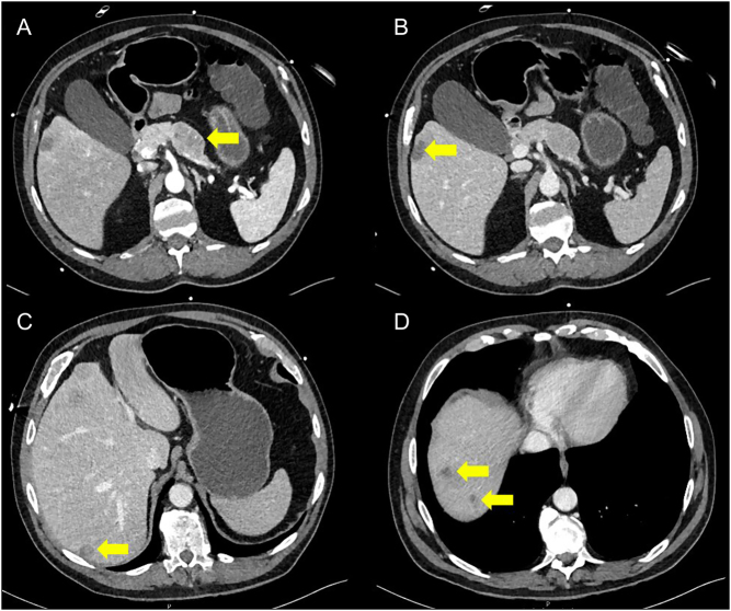
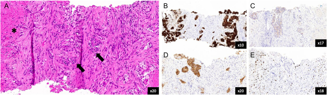
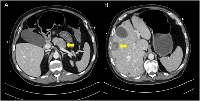
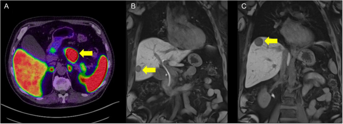
Summary: Vasoactive intestinal peptide-secreting tumours (VIPomas) are an extremely rare form of functional pancreatic neuroendocrine tumour with an estimated annual incidence of 1 in 10 million. Associated tumour hypersecretion of other peptides, including pancreatic polypeptide (PPomas), may also be seen. These malignancies classically present with a defined triad of refractory diarrhoea, hypokalaemia and metabolic acidosis known as Verner-Morrison syndrome. Diagnosis is frequently delayed, and the majority of patients will have metastatic disease at presentation. Symptoms are usually well controlled with somatostatin analogue administration. Here we report a case of metastatic mixed VIPoma/PPoma-induced diarrhoea causing renal failure so severe that ultrafiltration was required to recover adequate renal function.
Learning points: Profuse, watery diarrhoea is a common presenting complaint with a multitude of aetiologies. This, combined with the rarity of these tumours, makes diagnosis difficult and frequently delayed. A functional neuroendocrine tumour should be suspected when diarrhoea is unusually extreme, prolonged and common causes have been promptly excluded. These patients are likely to be profoundly unwell on presentation. They are extremely hypovolaemic with dangerous electrolyte and metabolic abnormalities. Aggressive initial rehydration and electrolyte replacement are imperative. A somatostatin analogue should be commenced as soon as the diagnosis is suspected. This is an extreme example of Verner-Morrison syndrome. We are unaware of another case where renal failure secondary to diarrhoea and dehydration was so severe that renal replacement therapy was required to restore adequate renal function, further emphasising how critically unwell these patients can be. Both the primary tumour and metastases showed a remarkably good and rapid response to somatostatin analogue administration. Cystic change and involution were noted on repeat imaging within days. Prior to his illness, this patient was extremely high functioning with no medical history. His diagnosis was an enormous psychological shock, and the consideration and care for his psychological well-being were a crucial part of his overall management. It highlights the importance of a holistic approach to cancer care and the role of the clinical nurse specialist within the cancer multidisciplinary team.
Figures
References
-
- Falconi M, Eriksson B, Kaltsas G, Bartsch DK, Capdevila J, Caplin M, Kos-Kudla B, Kwekkeboom D, Rindi G, Klöppel Get al.ENETS consensus guidelines update for the management of patients with functional pancreatic neuroendocrine tumors and non-functional pancreatic neuroendocrine tumors. Neuroendocrinology 2016103153–171. (10.1159/000443171) - DOI - PMC - PubMed
LinkOut - more resources
Full Text Sources
Molecular Biology Databases
