. 2022 Aug 24:11:e72435.
doi: 10.7554/eLife.72435.

Developmental stage-specific spontaneous activity contributes to callosal axon projections

Affiliations

Developmental stage-specific spontaneous activity contributes to callosal axon projections

Yuta Tezuka et al. Elife. .

Abstract

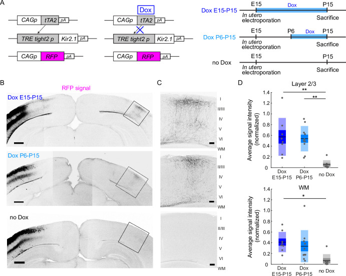
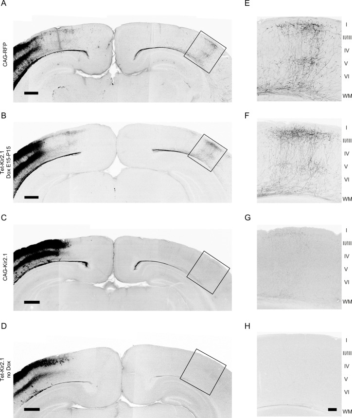
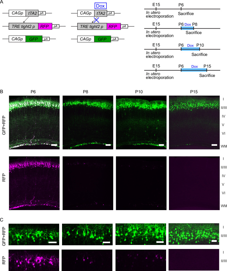
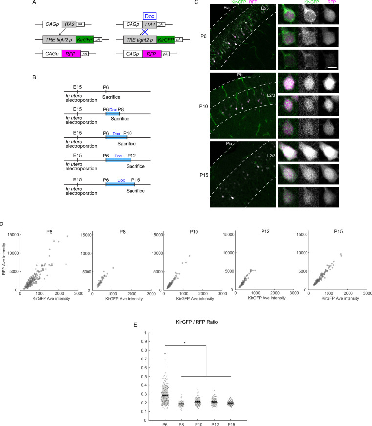
The developing neocortex exhibits spontaneous network activity with various synchrony levels, which has been implicated in the formation of cortical circuits. We previously reported that the development of callosal axon projections, one of the major long-range axonal projections in the brain, is activity dependent. However, what sort of activity and when activity is indispensable are not known. Here, using a genetic method to manipulate network activity in a stage-specific manner, we demonstrated that network activity contributes to callosal axon projections in the mouse visual cortex during a 'critical period': restoring neuronal activity during that period resumed the projections, whereas restoration after the period failed. Furthermore, in vivo Ca2+ imaging revealed that the projections could be established even without fully restoring highly synchronous activity. Overall, our findings suggest that spontaneous network activity is selectively required during a critical developmental time window for the formation of long-range axonal projections in the cortex.

Keywords: activity dependent; cerebral cortex; development; mouse; neuroscience.


Conflict of interest statement

YT, KH, KO, TH, YT No competing interests declared

Figures

Figure 1.
Figure 1.. Restoration of neuronal activity in the second postnatal week recovers callosal axon projections.
(A) Left, plasmids designed for temporally controlled expression of Kir2.1. Right, experimental timeline. Kir2.1 is a genetic tool to reduce neuronal activity. Transactivator tTA2s is expressed under the control of the CAG promoter (CAGp). Without doxycycline (Dox), tTA2s binds to the TRETight2 promoter (TRETight2p), and Kir2.1 expression is induced (left). With Dox administration, tTA2s cannot bind to the TRETight2 promoter, and Kir2.1 expression is suppressed (right). pA: polyA signal. (B) Coronal sections through the P15 cerebral cortex show the distribution of neurons expressing fluorescent proteins on the electroporated side and their axonal projections on the other side. Top: ‘Dox E15-P15’ condition where Dox was administered from E15 to P15; thus, Kir2.1 expression was suppressed throughout development. Middle: ‘Dox P6-15’ condition where Dox was administered from P6 to P15; thus, Kir2.1 expression was suppressed in the second postnatal week. Bottom: ‘no Dox’ condition where Kir2.1 is expressed throughout development. Scale bars, 500 μm. (C) Boxed regions in (B). WM: white matter. Scale bars, 100 μm. (D) Top, average signal intensity of fluorescently labeled callosal axons in layers 1–3. ‘Dox E15-P15’ group: n=8 sections from eight mice. ‘Dox P6-15’ group: n=10 sections from 10 mice. ‘no Dox’ group: n=7 sections from seven mice. **p<0.01 by Tukey–Kramer test. Bottom, average signal intensity of fluorescently labeled callosal axons in the white matter. *p<0.05 by Tukey–Kramer test.
Figure 1—figure supplement 1.
Figure 1—figure supplement 1.. Callosal axon projections with or without activity manipulation.
(A, E) Coronal sections through the P15 visual cortex show the distribution of neurons expressing fluorescent proteins on the electroporated side and their axonal projections on the other side. pCAG-TurboRFP was transfected to label callosal axons. n=10 sections from 10 mice. (B, F) TRETight2 promoter (pTRE-Tight2)-Kir2.1, pCAG-tTA2s, and pCAG-TurboRFP were transfected, and doxycycline (Dox) was administered from E15 to P15. Due to the presence of Dox, Kir2.1 expression is suppressed throughout development. n=8 sections from eight mice. (C, G) pCAG-Kir2.1 and pCAG-TurboRFP were transfected. Under the control of the pCAG, Kir2.1 is expressed throughout development. n=7 sections from seven mice. (D, H) pTRE-Tight2-Kir2.1, pCAG-tTA2s, and pCAG-TurboRFP were transfected, and Dox was not administered. Due to the absence of Dox, Kir2.1 is expressed throughout development. n = 7 sections from seven mice. Scale bars in A–D, 500 μm. Scale bar in H, 100 μm.
Figure 1—figure supplement 2.
Figure 1—figure supplement 2.. Validation of the Tet-off system with fluorescent proteins.
(A) pCAsal-EGFP, TRETight2 promoter (pTRE-Tight2)-TurboRFP, and CAG promoter (pCAG)-tTA2s were transfected, and doxycycline (Dox) was administered from P6. The blue bars indicate the period of Dox treatment. (B) Top panels show EGFP and TurboRFP signals. Bottom panels show TurboRFP signals. Note the rapid disappearance of TurboRFP signals after Dox administration. In (C), EGFP (top panels) and TurboRFP (bottom panels) signals in layer 2/3 are shown. Scale bars in (B), 100 μm. Scale bars in (C), 50 μm. P6, n=3 sections from three mice. P8, n=3 sections from three mice. P10, n=3 sections from three mice. P15, n=2 sections from two mice.
Figure 1—figure supplement 3.
Figure 1—figure supplement 3.. Validation of the Tet-off system with Kir2.1EGFP.
(A) CAG promoter (pCAG)-TurboRFP, TRETight2 promoter (pTRE-Tight2)-Kir2.1EGFP, and pCAG-tTA2s were transfected. (B) Doxycycline (Dox) was administered from P6. The blue bars indicate the period of Dox treatment. (C) Kir2.1EGFP (Kir-GFP) and TurboRFP (RFP) signals in P6, P10, and P15 cortical sections. Right panels show three representative neurons (in columns) with Kir-GFP and RFP signals (in rows) at each age. At P6 (before Dox treatment), the signals of Kir-GFP (stained with anti-GFP antibody) were primarily observed in the periphery of the soma and along dendrites, implying that Kir-GFP was transported to the cellular membrane. Note that RFP signals were localized in the central part of the cell body. At P10 and P15 (4 days and 9 days after Dox treatment), Kir-GFP signals were not preferentially observed in the periphery of the soma and along dendrites anymore. Low-level signals in the central part of the cell body may suggest low-level expression of Kir-GFP even after Dox treatment. Alternatively, these may reflect the contaminated RFP signals in the acquisition of the GFP channel. (D) Average signal intensity of Kir-GFP (x-axis) and RFP (y-axis) in each cell is displayed at P6, P8, P10, P12, and P15. (E) Each dot represents KirGFP/RFP ratio of a cell at each age. Note that KirGFP/RFP ratio decreases after Dox administration at P6. Scale bar in the left panel of (C), 50 μm; right panel, 10 μm. P6, n=10 sections from three mice. P8, n=7 sections from two mice. P10, n=10 sections from three mice. P12, n=10 sections from three mice. P15, n=10 sections from three mice.
Figure 1—figure supplement 4.
Figure 1—figure supplement 4.. The width and laminar distribution of callosal axon projections.
(A, B) The width of callosal axon innervation zone in layers 2/3 (A) and 5 (B). The width of the zone along the mediolateral axis of the cortex was measured. Although callosal axon innervation zone was present in both layers 2/3 and 5 of doxycycline (Dox) P6-15 mice, the extent was not as much as in Dox E15-P15 mice, suggesting that some aspects of callosal projections might be partially recovered in Dox P6-15 mice. ‘Dox E15-P15’ group: n=8 sections from eight mice. ‘Dox P6-15’ group: n=9 sections from nine mice. ‘no Dox’ group: n=7 sections from seven mice. ‘RFP’ group: n=7 sections from seven mice. (C) Densitometric line scans of fluorescence intensity across all cortical layers. ‘Dox E15-P15’ group: n=8 sections from eight mice. ‘Dox P6-15’ group: n=9 sections from nine mice. ‘no Dox’ group: n=7 sections from seven mice. ‘RFP’ group: n=7 sections from seven mice.
Figure 2.
Figure 2.. Effect of activity restoration on callosal axon projections using designer receptors exclusively activated by designer drugs (DREADD).
(A) Experimental design and timeline. Kir2.1 expression and hM3DGq expression plasmids were transfected at E15, clozapine-N-oxide (CNO) or saline was injected daily from P10 to P14, and brains were fixed at P15. (B) Fluorescently labeled callosal axon projections in the P15 cortex. Scale bar, 100 μm. (C) Average signal intensity of fluorescently labeled callosal axons in layers 1–3. ‘Kir + hM3 DGq with CNO injections’ group; n=11 sections from 11 mice. ‘Kir + hM3 DGq with saline injections’ group; n=10 sections from 10 mice. ‘Kir with CNO injections’ group: n=4 sections from four mice. ‘RFP‘ group (CAG promoter [pCAG]-TurboRFP was electroporated, no injection was performed): n=10 sections from 10 mice. ‘Kir’ group (pCAG-Kir2.1 was electroporated, no injection was performed): n=7 sections from seven mice. ***p<0.001 by Tukey–Kramer test.
Figure 3.
Figure 3.. A critical period for restoration of callosal axon projections.
(A) Experimental design and timeline. CAG promoter (pCAG)-tTA2s and TRETight2 promoter (pTRETight2)-Kir2.1 plasmids were transfected at E15, and doxycycline (Dox) was administered from P6 to P15, from P9 to P18, or from P12 to P21. (B) Fluorescently labeled callosal axon projections in the P15 cortex. Scale bar, 100 μm. (C) Average signal intensity of fluorescently labeled callosal axons in layers 1–3. ‘Dox P6-15’ group: n=10 sections from 10 mice. ‘Dox P9-P18’ group: n=9 sections from nine mice. ‘Dox P12-P21’ group: n=10 sections from 10 mice. ***p<0.001 by Tukey–Kramer test.
Figure 4.
Figure 4.. Partial restoration of synchronous activity revealed by in vivo two-photon Ca2+ imaging.
(A) A representative image of OBG1-loaded (gray) and FP635-expressing (magenta) neurons in the P13 mouse visual cortex obtained by two-photon microscopy. Scale bar: 50 μm. Numbers shown in the image correspond to the cell numbers shown in panel (B). (B) Ca2+ traces from representative FP635+ (#1–4) and FP635− (#5–10) neurons. Obtained from the same recording session as shown in the bottom panel. (C) Top: an active period plot of all identified FP635+ (magenta) and FP635− (black) neurons in the top image. Bottom: corresponding participation rate. Events with a participation rate above 60% were regarded as H events. (D) A representative active period plot and corresponding participation rate from a Tet-Kir2.1 with doxycycline (Dox) mouse. Because of Dox treatment from P6, Kir2.1 expression was suppressed from P6: Kir OFF. (E) A representative active period plot and corresponding participation rate from a Tet-Kir2.1 without Dox mouse. Because of the absence of Dox, Kir2.1 was expressed throughout development: Kir ON. (F) Histograms of synchronous activity frequency with different participation rates from FP635-electroporated (Left: FP only), Tet-Kir2.1 with Dox (Middle: Kir OFF Dox P6), and Tet-Kir2.1 without Dox (Right: Kir ON) mice. (G) Quantifications of H (>60%) and L (20–60%) events. FP only, five mice. Kir OFF (Dox P6), five mice. Kir ON, five mice. *p<0.05, **p<0.01, ***p<0.001 by Tukey–Kramer test.

Similar articles

Cited by

References

    1. Ackman JB, Burbridge TJ, Crair MC. Retinal waves coordinate patterned activity throughout the developing visual system. Nature. 2012;490:219–225. doi: 10.1038/nature11529. - DOI - PMC - PubMed
    1. Ackman JB, Crair MC. Role of emergent neural activity in visual map development. Current Opinion in Neurobiology. 2014;24:166–175. doi: 10.1016/j.conb.2013.11.011. - DOI - PMC - PubMed
    1. Alexander GM, Rogan SC, Abbas AI, Armbruster BN, Pei Y, Allen JA, Nonneman RJ, Hartmann J, Moy SS, Nicolelis MA, McNamara JO, Roth BL. Remote control of neuronal activity in transgenic mice expressing evolved G protein-coupled receptors. Neuron. 2009;63:27–39. doi: 10.1016/j.neuron.2009.06.014. - DOI - PMC - PubMed
    1. Allène C, Cattani A, Ackman JB, Bonifazi P, Aniksztejn L, Ben-Ari Y, Cossart R. Sequential generation of two distinct synapse-driven network patterns in developing neocortex. The Journal of Neuroscience. 2008;28:12851–12863. doi: 10.1523/JNEUROSCI.3733-08.2008. - DOI - PMC - PubMed
    1. Blankenship AG, Feller MB. Mechanisms underlying spontaneous patterned activity in developing neural circuits. Nature Reviews. Neuroscience. 2010;11:18–29. doi: 10.1038/nrn2759. - DOI - PMC - PubMed

Publication types