. 2022 Dec;18(4):469-479.
doi: 10.1007/s11302-022-09894-y. Epub 2022 Aug 24.

P2X7-mediated alteration of membrane fluidity is associated with the late stages of age-related macular degeneration

Affiliations

P2X7-mediated alteration of membrane fluidity is associated with the late stages of age-related macular degeneration

Candace Drysdale et al. Purinergic Signal. 2022 Dec.

Abstract

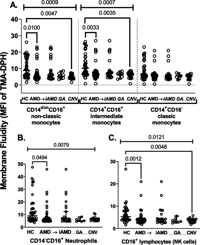
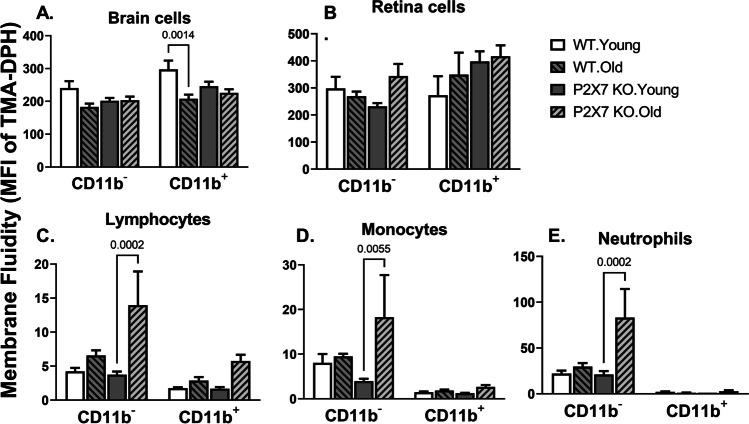
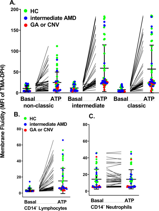
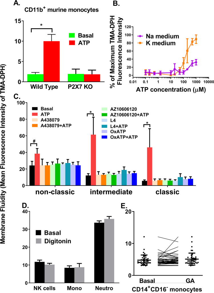
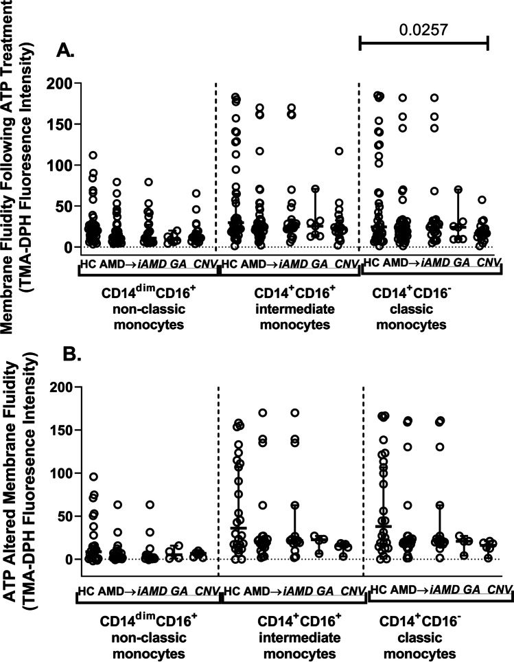
We have shown deficits in monocyte phagocytosis from patients with age-related macular degeneration (AMD). Cell membrane fluidity is known to affect phagocytic capacity and leucocyte functionality more generally. Therefore, we examined membrane fluidity of peripheral blood leucocytes in human patients with AMD and in the P2X7 null mouse model of AMD using flow cytometry with a fluorescent probe for fluidity, TMA-DPH. The results showed that membrane fluidity was decreased in all leucocyte types of late AMD relative to healthy controls (HC) including monocytes, neutrophils and lymphocytes but this was not apparent in earlier stages of AMD. Further analysis of factors contributing to membrane fluidity indicated that pre-treatment of monocytes and lymphocytes with ATP greatly increased membrane fluidity in humans and mice. Evidence from P2X7 null mice and P2X7 antagonists confirmed that these ATP-driven increases in membrane fluidity were mediated by P2X7 but were not associated with the classic P2X7 functions of pore formation or phagocytosis. Analysis of P2X7 expression indicated that receptor levels were elevated in classic monocytes of late AMD patients, further suggesting the P2X7 may contribute to altered plasma membrane properties. Our findings identified a novel biological function of P2X7 in modulating membrane fluidity of leucocytes and demonstrated reduced membrane fluidity in cellular changes associated with the late stage of AMD.

Keywords: ATP; Age-related macular degeneration; Membrane fluidity; Monocyte; P2X7; TMA-DPH.


Conflict of interest statement

The authors declare no competing interests.

Figures

Fig. 1
Fig. 1
Membrane fluidity of human peripheral leukocytes. Peripheral blood from healthy controls (n = 40) and AMD patients (n = 49, including 22 iAMD, 7 GA and 22 CNV) was labelled with CD14 and CD16 and incubated with TMA-DPH for 5 min prior to lysing. The log mean fluorescence intensity (MFI) of TMA-DPH was measured by flow cytometry with a violet laser (405 nm). Cells were gated according to forward/side scatter and CD14/CD16 expression as A monocytes, B neutrophils and C NK cells. Results are presented as median ± 95% CI. A Kruskal–Wallis test with Dunn’s multiple comparisons was used for analysis. P values of all groups and individual group comparison are indicated
Fig. 2
Fig. 2
Membrane fluidity of murine cells. Cells were collected from the A brain, B retina and CE peripheral circulation of wild type C57BL/6 mice (aged 2–4 months and 20–22 months, n = 7 and 6 respectively) and P2X7 knockout mice (aged 2–4 months and 20–22 months, n = 6 and 5 respectively). Cells were labelled with APC-conjugated anti-mouse CD11b antibody and then stained with TMA-DPH for 5 min. Cells were gated according to forward/side scatter and CD11b expression as A brain cells, B retina cells, C lymphocytes, D monocytes and E neutrophils. Results are presented as mean ± SED. Two-way ANOVA with Tukey’s multiple comparison was used for analysis. P values of individual group comparison are indicated
Fig. 3
Fig. 3
ATP increases membrane fluidity in monocytes and lymphocytes but not neutrophils. Peripheral blood from healthy controls (n = 25), intermediate AMD (n = 11) and advanced AMD (GA or CNV, n = 15) was incubated with or without 1 mM ATP for 5 min at 37 °C, followed by incubation with 10 µM TMA-DPH for 5 min. Red cells were lysed and leukocytes were labelled with fluorescent anti-CD14 and anti-CD16 mAb. Cells were gated according to forward/side scatter and CD14/CD16 expression as A monocytes, B lymphocytes and C neutrophils. P < 0.0001 for all paired comparison of basal vs. ATP in three subsets of monocytes and in lymphocytes. A, B, Wilcoxon test
Fig. 4
Fig. 4
ATP-stimulated membrane fluidity is via activation of the P2X7 receptor but independent of pore formation or phagocytosis. A Peripheral blood collected from wild type or P2X7 knockout C57BL mice was labelled with CD11b and then pre-treated with or without ATP prior to the addition of TMA-DPH. Blood was then lysed. Results are presented as mean ± SD (n = 6). B Human PBMCs (1 × 107/mL) were resuspended in 200-µL Na medium or K medium and treated with various concentrations of ATP for 5 min before the addition of 10 µM TMA-DPH. Cells were then stained with APC-conjugated anti-CD14 mAb and gated on CD14+ monocyte population. Results are presented as mean ± SD (n = 3). C PBMCs were resuspended in 200-µL K medium and treated with P2X7 antagonists A438079 (1 µM), AZ10606120 (1 µM), anti-P2X7 mAb (clone L4, 100 µg/mL) or oxidized ATP (100 µM) prior to the addition of ATP and TMA-DPH. Results are presented as mean ± SD (n = 6). D Whole blood was incubated with or without 0.0015% digitonin (a pore-forming detergent) for 5 min prior to the addition of 10 µM TMA-DPH. Leukocytes are gated on CD14CD16+ lymphocytes (NK cells), CD14+ monocytes and CD14CD16+ neutrophils. Results are presented as mean ± SD (n = 3). E Peripheral blood mononuclear cells (PBMCs) were labelled with CD14 and CD16 then incubated with 100 µg/mL CPX for 5 min at 37 °C prior to the addition of 10 µM TMA-DPH. Blood was lysed and washed, followed by CD14 and CD16 labelling (n = 44). *P < 0.001; #P = 0.0120 (paired Wilcoxon test)
Fig. 5
Fig. 5
ATP increased membrane fluidity of healthy controls and AMD participants. Peripheral blood from healthy controls (n = 39) and AMD participants (n = 77, including 50 early stages AMD, 7 GA and 20 CNV) were labelled with CD14 and CD16 then pre-treated with 1 mM ATP prior to the addition of TMA-DPH. A TMA-DPH MFI following ATP treatment in gated monocyte subsets. B ATP-promoted membrane fluidity changes were calculated as membrane fluidity on ATP-treated cells minus untreated cells. Results are presented as median ± 95% CI. Kruskal–Wallis test with Dunn’s multiple comparisons was used for analysis
Fig. 6
Fig. 6
P2X7 surface expression in monocyte subsets. Peripheral blood from healthy controls (n = 56) and AMD participants (n = 65 including 40 early stages AMD, 11 GA and 14 CNV) was stained with Alexa647-conjugated anti-human P2X7 mAb (clone L4), FITC-CD16, PE-CD4 and PerCP-CD14 mAb prior to lysis and washing. Cells were gated according to forward/side scatter and CD14/CD16/CD4 expression as A three subsets of monocytes as stratified by CD14/CD16 expression, B total CD14+ monocytes and C CD14+CD4+ monocytes. The log mean fluorescence intensity of Alexa647-P2X7 was measured by flow cytometry in gated monocyte subpopulations. Results are presented as median ± 95% CI. Kruskal–Wallis test with Dunn’s multiple comparisons was used for analysis

Similar articles

Cited by

References

    1. Fleckenstein M, Keenan TDL, Guymer RH, et al. Age-related macular degeneration. Nat Rev Dis Primers. 2021;7(1):31. doi: 10.1038/s41572-021-00265-2. - DOI - PubMed
    1. Wu Z, Guymer RH. Can the onset of atrophic age-related macular degeneration be an acceptable endpoint for preventative trials? Ophthalmologica. 2020;243(6):399–403. doi: 10.1159/000510887. - DOI - PubMed
    1. Gu BJ, Huang X, Avula PK, et al. Deficits in monocyte function in age related macular degeneration: a novel systemic change associated with the disease. Front Med. 2021;8:634177. doi: 10.3389/fmed.2021.634177. - DOI - PMC - PubMed
    1. Sluyter R, Shemon AN, Hughes WE, et al. Canine erythrocytes express the P2X7 receptor: greatly increased function compared with human erythrocytes. Am J Physiol Regul Integr Comp Physiol. 2007;293(5):R2090–2098. doi: 10.1152/ajpregu.00166.2007. - DOI - PubMed
    1. Wong J, Gu BJ, Teoh H, et al. Flow cytometry identifies an early stage of platelet apoptosis produced by agonists of the P2X1 and P2X7 receptors. Platelets. 2022;33(4):1–11. doi: 10.1080/09537104.2021.1981844. - DOI - PubMed

Publication types

Substances