. 2022 Sep 14;145(9):2982-2990.
doi: 10.1093/brain/awac199.

Aqp4 stop codon readthrough facilitates amyloid-β clearance from the brain

Affiliations

Aqp4 stop codon readthrough facilitates amyloid-β clearance from the brain

Darshan Sapkota et al. Brain. .

Abstract

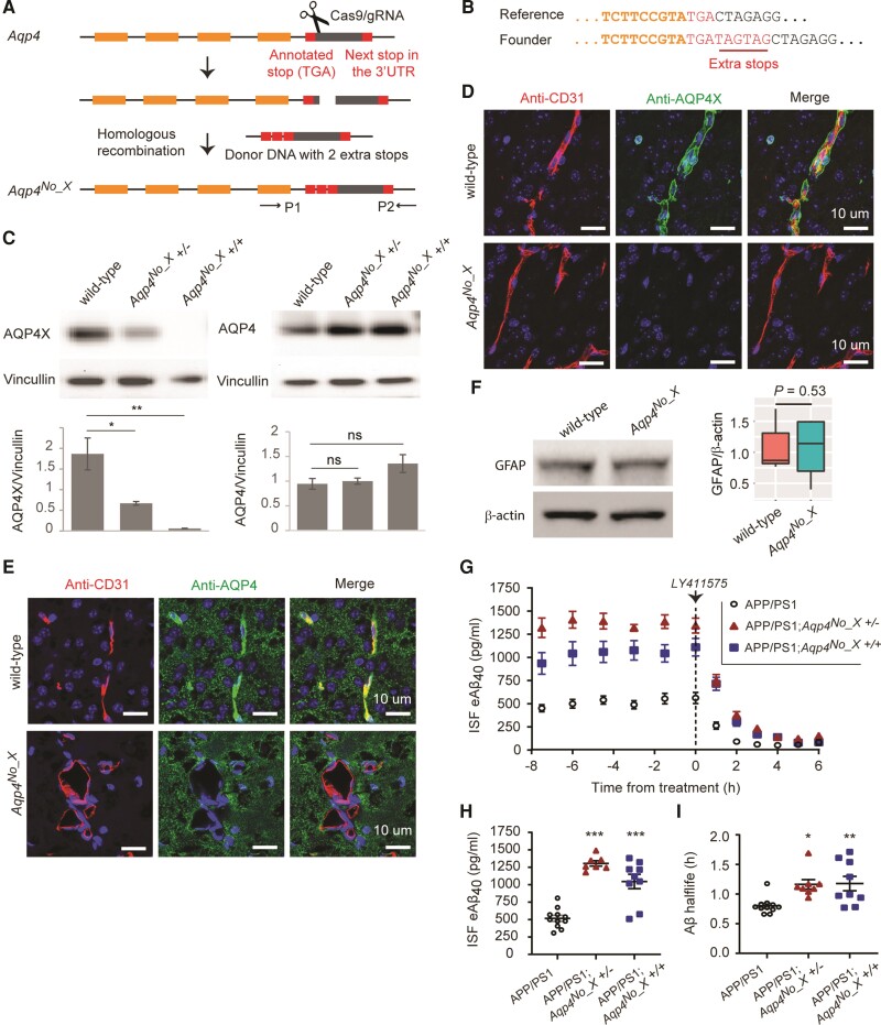
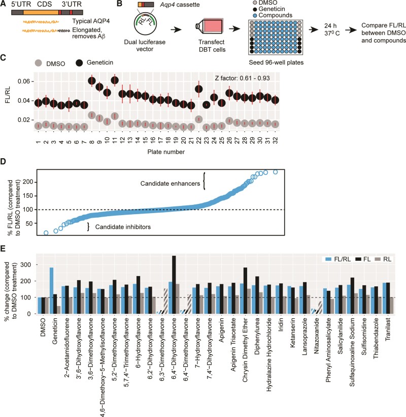
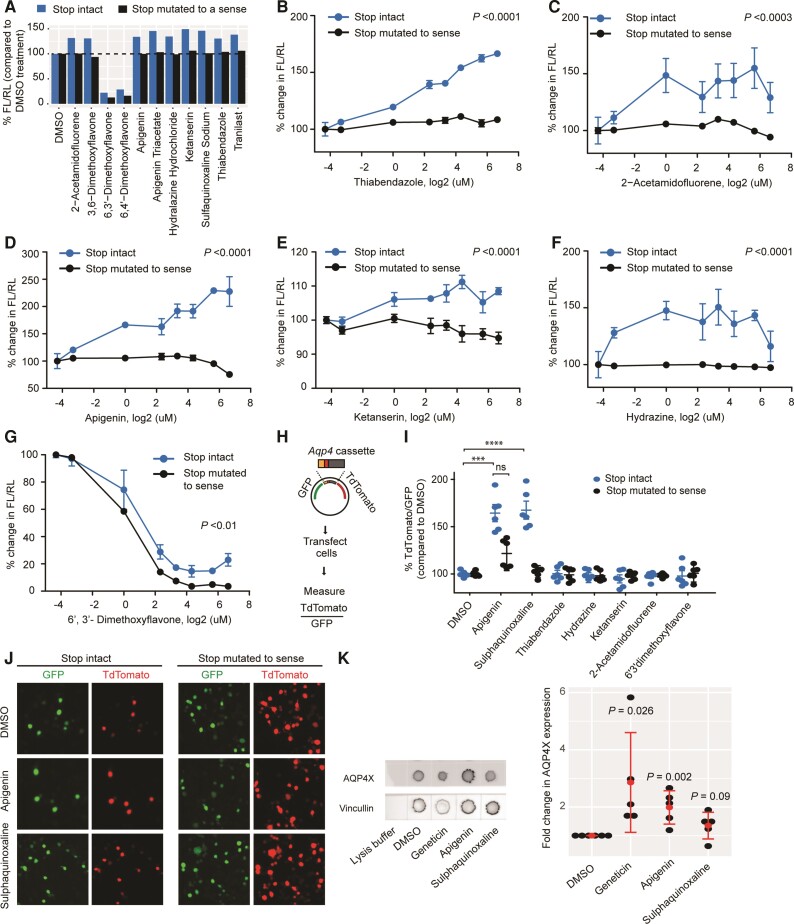
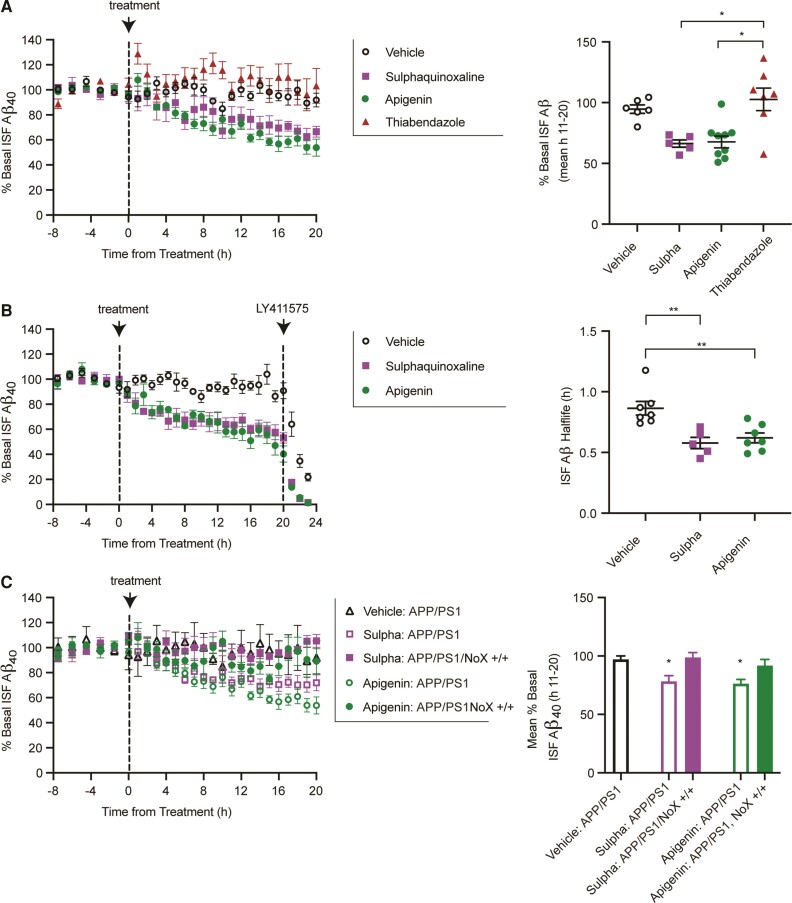
Alzheimer's disease is initiated by the toxic aggregation of amyloid-β. Immunotherapeutics aimed at reducing amyloid beta are in clinical trials but with very limited success to date. Identification of orthogonal approaches for clearing amyloid beta may complement these approaches for treating Alzheimer's disease. In the brain, the astrocytic water channel Aquaporin 4 is involved in clearance of amyloid beta, and the fraction of Aquaporin 4 found perivascularly is decreased in Alzheimer's disease. Further, an unusual stop codon readthrough event generates a conserved C-terminally elongated variant of Aquaporin 4 (AQP4X), which is exclusively perivascular. However, it is unclear whether the AQP4X variant specifically mediates amyloid beta clearance. Here, using Aquaporin 4 readthrough-specific knockout mice that still express normal Aquaporin 4, we determine that this isoform indeed mediates amyloid beta clearance. Further, with high-throughput screening and counterscreening, we identify small molecule compounds that enhance readthrough of the Aquaporin 4 sequence and validate a subset on endogenous astrocyte Aquaporin 4. Finally, we demonstrate these compounds enhance brain amyloid-β clearance in vivo, which depends on AQP4X. This suggests derivatives of these compounds may provide a viable pharmaceutical approach to enhance clearance of amyloid beta and potentially other aggregating proteins in neurodegenerative disease.

Keywords: Alzheimer’s disease; Aqp4; amyloid beta; glymphatic; readthrough.


Conflict of interest statement

The authors report no competing interests.

Figures

Figure 1
Figure 1
AQPX regulates clearance of Aβ. (A) CRISPR–Cas9 for generating the Aqp4No_X mouse. A double-strand break is made using a gRNA and two extra stop codons are introduced using a donor oligo for homologous recombination. P1 and P2 indicate primers designed to screen the founders. (B) Genotyping of founder mice. Illumina sequencing read from a founder is shown. (C) Western blot to validate mice. Note that anti-AQP4 recognizes both normal and readthrough AQP4s and hence continues to generate signal in the Aqp4No_X mouse. n = 3, t-test. (D and E) Characterization of Aqp4No_X mice. Perivascular AQP4, i.e. AQP4X, is lost while parenchymal AQP4 is intact in mutant mice. CD31 marks endothelial cells. Blue = nuclear staining with DAPI. (F) No gliosis is observed in mutant mice, as apparent from GFAP western blot. n = 3, t-test. (G) Steady-state exchangeable hippocampal ISF Aβ (eAβ) was measured longitudinally using in vivo microdialysis for 7.5 h followed by administration of a γ-secretase inhibitor (LY411575, 3 mg/kg intraperitoneally) then Aβ measured hourly for an additional 6 h. (H) Steady-state ISF eAβ concentration in APP/PS1 Aqp4 wild-type mice is 516.0 ± 42.8 pg/ml and significantly elevated to 1305 ± 38.82 pg/ml and 1045 ± 102.9 in Aqp4No_X +/− and +/+, respectively. Concentrations are not significantly different between +/− and +/+. (I) ISF eAβ elimination half-life in APP/PS1 Aqp4 wild-type mice is 0.80 + 0.04 h and significantly prolonged to 1.16 ± 0.08 and 1.18 ± 0.12 h in Aqp4No_X +/− and +/+ mice, respectively. Data presented as mean ± SEM. See Supplementary Table 1 for n and statistical tests. See Supplementary Fig. 4 for full images of blots used to prepare C and F. ***P ≤ 0.0001; **P ≤ 0.01; *P ≤ 0.05.
Figure 2
Figure 2
High-throughput screening identifies readthrough modulators. (A) Schematic illustration of Aqp4 readthrough. (B) Screening procedure. Transfected cells were seeded into wells pre-spotted with compounds, DMSO (no-treatment control) or Geneticin (positive control). The last column received control cells in which the Aqp4 stop in the vector was mutated to a sense codon. (C) Separation bands between the no-treatment and positive controls and a summary of Z-factors for the plates used in screening. Dots and error bars represent means and 3 SD, respectively. (D) Quantile analysis of the screening results. (E) Compounds enhancing (solid bars) or inhibiting (patterned bars) FL/RL values by at least 50%. The change in FL/RL is largely due to the modulation of FL, not RL.
Figure 3
Figure 3
Validation of candidate compounds. (A) Retesting with dual-luciferase vectors in which the Aqp4 stop codon is either intact (blue) or mutated to a sense codon (black). Compounds altering readthrough by at least 30% are shown. Enhancers cease activity in the absence of stop codon. (BG) Dose-response curves for candidate compounds. Concentrations are log2-transformed. For log transformation of 0 µM (DMSO alone), a value of 0.05 µM was used. Enhancers cease to be active (BF), whereas an inhibitor continues to be active (G) in the absence of stop codon. n = 3. ANOVA followed by a post hoc Tukey was used to compare the means for the two constructs across concentrations. (H) Dual-fluorescence assay. Readthrough is measured as TdTomato/GFP (I) dual-fluorescence assays in which the Aqp4 stop codon is either intact (blue) or mutated to a sense codon (black). ****P ≤ 0.0001; ***P ≤ 0.001, n = 3, t-test. (J) Representative images for dual-fluorescence assay with apigenin and sulphaquinoxaline. (K) Dot blot demonstrates apigenin enhances readthrough of endogenous Aqp4 transcript in cultured astrocytes. AQP4X expression is normalized to Vincullin. Red dots represent means and bars represents standard deviations. t-test comparing DMSO and compounds. n = 3 **P ≤ 0.01; *P ≤ 0.05.
Figure 4
Figure 4
Compounds lower Aβ in a readthrough dependent manner. (A) Continuous reverse microdialysis administration of sulphaquinoxaline (200 μM) and apigenin (100 μM) reduces hippocampal ISF Aβ in APP/PS1 mice over a 20 h period, while a non-readthrough promoting compound, thiabendazole (400 μM), does not (left). Concentrations vary based on in vitro potency. Sulphaquinoxaline and apigenin reduce ISF Aβ by 29.9 ± 3.2 and 27.3 ± 5.2%, respectively, compared to vehicle-treated mice (n = 6) (right). In contrast, thiabendazole was not significantly different from vehicle, but significantly higher than the other treatment groups (mean over hours 11–20 of administration; Kruskal–Wallis test). (B) A separate cohort of APP/PS1 mice was treated with apigenin, sulphaquinoxaline or vehicle by reverse microdialysis for 20 h following by LY411575 (3 mg/kg intraperitoneally). The ISF Aβ elimination half-lives in apigenin and sulphaquinoxaline treated mice were 0.62 ± 0.04 and 0.57 ± 0.47 h, respectively, compared to vehicle-treated mice at 0.86 ± 0.56 h. (C) Sulphaquinoxaline and apigenin do not alter interstitial Aβ in APP/PS1 mice deficient in Aqp4 readthrough. Data presented as mean ± SEM. See Supplementary Table 1 for n and statistical tests. *P = 0.01, **P = 0.001.

Comment in

References

    1. Iliff JJ, Wang M, Liao Y, et al. A paravascular pathway facilitates CSF flow through the brain parenchyma and the clearance of interstitial solutes, including amyloid β. Sci Transl Med. 2012;4:147ra111. - PMC - PubMed
    1. Mestre H, Hablitz LM, Xavier AL, et al. Aquaporin-4-dependent glymphatic solute transport in the rodent brain. eLife. 2018;7:e40070. - PMC - PubMed
    1. Xu Z, Xiao N, Chen Y, et al. Deletion of aquaporin-4 in APP/PS1 mice exacerbates brain Aβ accumulation and memory deficits. Mol Neurodegener. 2015;10:58. - PMC - PubMed
    1. Alkalaeva EZ, Pisarev AV, Frolova LY, Kisselev LL, Pestova TV. In vitro reconstitution of eukaryotic translation reveals cooperativity between release factors eRF1 and eRF3. Cell. 2006;125:1125–1136. - PubMed
    1. Firth AE, Brierley I. Non-canonical translation in RNA viruses. J Gen Virol. 2012;93:1385–1409. - PMC - PubMed

Publication types