. 2022 Aug 24;17(8):e0273542.
doi: 10.1371/journal.pone.0273542. eCollection 2022.

Knockdown of CD146 promotes endothelial-to-mesenchymal transition via Wnt/β-catenin pathway

Affiliations

Knockdown of CD146 promotes endothelial-to-mesenchymal transition via Wnt/β-catenin pathway

Zhao-Yu Zhang et al. PLoS One. .

Abstract

Purpose: Cardiac fibrosis is characterized by the excessive deposition of extracellular matrix (ECM) proteins and leads to the maladaptive changes in myocardium. Endothelial cells (ECs) undergoing mesenchymal transition contributes to the occurrence and development of cardiac fibrosis. CD146 is an adhesion molecule highly expressed in ECs. The present study was performed to explore the role of CD146 in modulating endothelial to mesenchymal transition (EndMT).

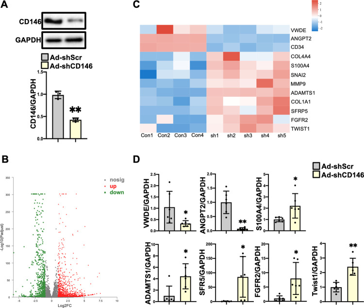
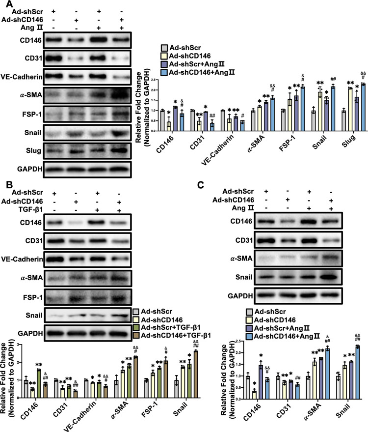
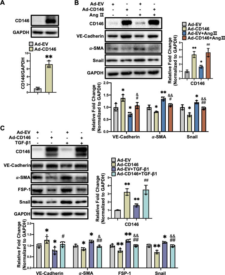
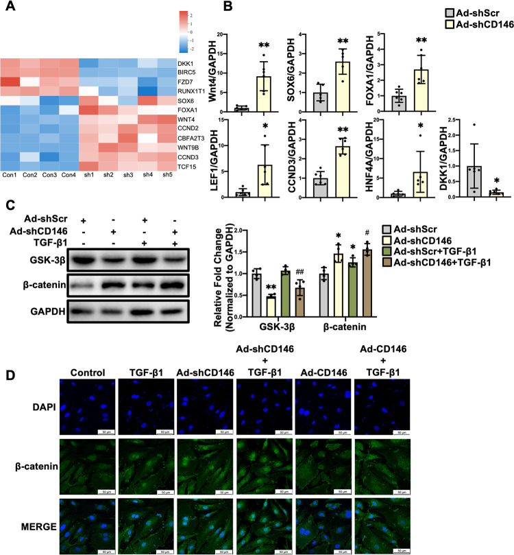
Methods: C57BL/6 mice were subjected to subcutaneous implantation of osmotic minipump infused with angiotensin II (Ang Ⅱ). Adenovirus carrying CD146 short hairpin RNA (shRNA) or CD146 encoding sequence were infected into cultured human umbilical vein endothelial cells (HUVECs) followed by stimulation with Ang II or transforming growth factor-β1 (TGF-β1). Differentially expressed genes were revealed by RNA-sequencing (RNA-Seq) analysis. Gene expression was measured by quantitative real-time PCR, and protein expression and distribution were determined by Western blot and immunofluorescence staining, respectively.

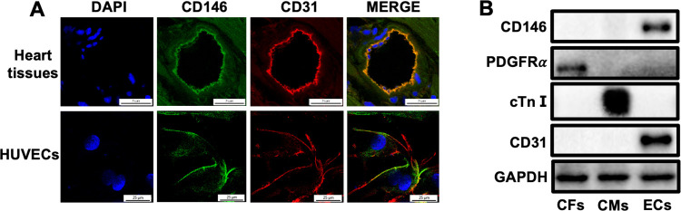
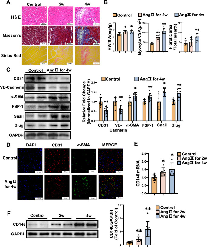
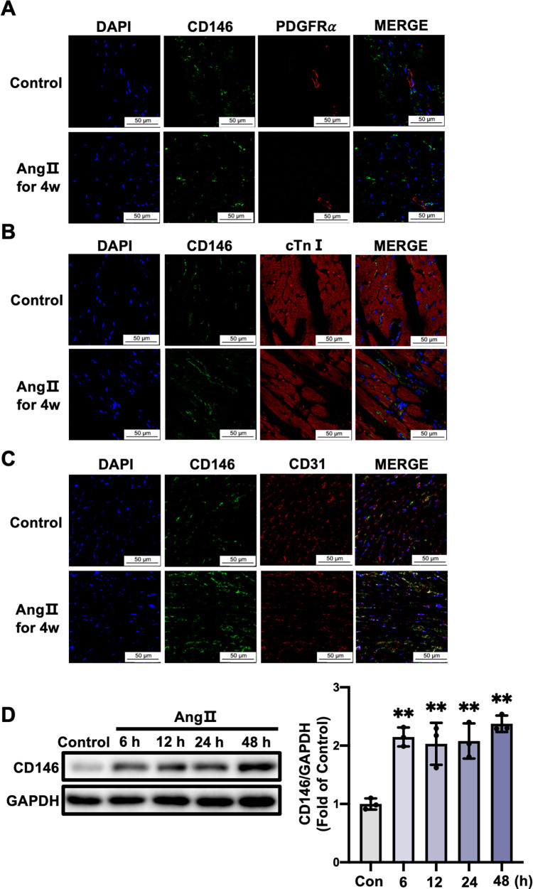
Results: CD146 was predominantly expressed by ECs in normal mouse hearts. CD146 was upregulated in ECs but not fibroblasts and myocytes in hearts of Ang II-infused mice and in HUVECs stimulated with Ang Ⅱ. RNA-Seq analysis revealed the differentially expressed genes related to EndMT and Wnt/β-catenin signaling pathway. CD146 knockdown and overexpression facilitated and attenuated, respectively, EndMT induced by Ang II or TGF-β1. CD146 knockdown upregulated Wnt pathway-related genes including Wnt4, LEF1, HNF4A, FOXA1, SOX6, and CCND3, and increased the protein level and nuclear translocation of β-catenin.

Conclusions: Knockdown of CD146 exerts promotional effects on EndMT via activating Wnt/β-catenin pathway and the upregulation of CD146 might play a protective role against EndMT and cardiac fibrosis.


Conflict of interest statement

The authors have declared that no competing interests exist.

Figures

Fig 1
Fig 1. CD146 is predominantly expressed in ECs of adult mouse hearts.
A CD146 expression in adult mouse hearts and HUVECs was determined by immunofluorescence staining (scale bars indicate 25 μm). Green represents CD146; red represents CD31; blue represents nuclei. B CD146 protein expression in CFs, CMs and ECs isolated from adult mouse hearts was detected by Western blot analysis. GAPDH served as an internal control. CFs: cardiac fibroblasts, CMs: cardiac myocytes, ECs: endothelial cells, cTnⅠ: cardiac troponin Ⅰ.
Fig 2
Fig 2. CD146 is upregulated in the fibrotic mouse hearts.
A Mice were subjected to Ang Ⅱ (1.4 mg/kg/day) infusion for 2 and 4 weeks. Representative images of H&E staining, Masson’s trichrome staining, and Sirius Red staining (scale bars indicate 100 μm). B The ratio of heart weight to body weight (HW/BW) of mice was calculated. Myocyte cross–sectional area (CSA) and connective tissue percentage were measured from images captured from H&E–stained and Masson’s trichrome–stained sections, respectively. C The protein levels of CD31, VE–Cadherin, α–SMA, FSP–1, Snail, and Slug in left ventricles from control and Ang Ⅱ–infusion groups for 4 weeks were measured by Western blot analysis. GAPDH served as an internal control. D Representative double–immunofluorescence images of CD31 and α–SMA expression in left ventricles from control and Ang Ⅱ–infusion groups for 4 weeks (scale bars indicate 75 μm). Red represents CD31; green represents α–SMA; blue represents nuclei. Levels of CD146 mRNA (E) and protein (F) in left ventricles from control, Ang Ⅱ for 2 weeks, and Ang Ⅱ for 4 weeks. GAPDH served as an internal control. *P<0.05, **P<0.01 vs. Control.
Fig 3
Fig 3. CD146 upregulation by Ang II is mainly restricted to cardiac ECs.
Representative double–immunofluorescence images of CD146 with PDGFRα (A), cTnⅠ (B) and CD31 (C) in left ventricles from control and 4–week Ang II–infused mice (scale bars indicate 50 μm). Green represents CD146; red represents PDGFRα, cTnⅠ and CD31, respectively; blue represents nuclei. D HUVECs were treated with Ang Ⅱ (1×10⁻⁶ mol/L) for the indicated times and the protein levels of CD146 were measured by Western blot analysis. GAPDH served as an internal control. *P<0.05, **P<0.01 vs. Control.
Fig 4
Fig 4. ECs with CD146 knockdown acquire a mesenchymal phenotype.
A HUVECs were infected with Ad–shScr and Ad–shCD146 for 48 h. The protein level of CD146 was measured by Western blot analysis. GAPDH served as an internal control. B Numbers of differentially expressed genes in comparison of HUVECs infected with Ad–shScr and Ad–shCD146. C Heatmap depicting the differentially expressed genes related to EndMT. D The mRNA levels of VWDE, ANGPT2, S100A4, ADAMTS1, SFR5, FGFR2, and Twist1 were measured by qRT–PCR analysis. *P<0.05, **P<0.01 vs. Ad–shScr. Ad–shScr: Ad–shScramble.
Fig 5
Fig 5. Knockdown of CD146 promotes Ang Ⅱ–and TGF–β1–induced EndMT.
HUVECs were infected with Ad–shScr or Ad–shCD146 for 48 h followed by Ang Ⅱ (1×10⁻⁶ mol/L) (A) or TGF–β1 (10 ng/ml) (B) treatment for 48 h. The protein levels of CD146, CD31, VE–Cadherin, α–SMA, FSP–1, Snail and Slug were measured by Western blot analysis. GAPDH served as an internal control. C MCMECs were infected with Ad–shScr or Ad–shCD146 for 48 h followed by Ang Ⅱ (1×10⁻⁶ mol/L) treatment for 48 h. The protein levels of CD146, CD31, α–SMA and Snail were measured by Western blot analysis. GAPDH served as an internal control. *P<0.05, **P<0.01 vs. Ad–shScr, #P<0.05, ## P<0.01 vs. Ad–shScr+Ang Ⅱ or TGF–β1. &P<0.05, && P<0.01 vs. Ad–shCD146. Ad–shScr: Ad–shScramble.
Fig 6
Fig 6. Overexpression of CD146 suppresses Ang Ⅱ–and TGF–β1–induced EndMT in HUVECs.
A HUVECs were infected with Ad–EV and Ad–CD146 for 48 h. The protein level of CD146 was measured by Western blot analysis. GAPDH served as an internal control. HUVECs were infected with Ad–EV or Ad–CD146 for 48h followed by Ang Ⅱ (1×10⁻⁶ mol/L) (B) or TGF–β1 (10 ng/ml) (C) treatment for 48h. The protein levels of CD146, VE–Cadherin, α–SMA, FSP–1, and Snail were measured by Western blot analysis. GAPDH served as an internal control. *P<0.05, **P<0.01 vs. Ad–EV, #P<0.05, ## P<0.01 vs. Ad–EV+ Ang Ⅱ or TGF–β1. &P<0.05, &&P<0.01 vs. Ad–EV. Ad–EV: Ad–empty vector.
Fig 7
Fig 7. Knockdown of CD146 leads to the activation of canonical Wnt/β–catenin pathway in HUVECs.
A Heatmap depicting the differentially expressed genes related to Wnt signaling pathway. B The mRNA levels of Wnt4, SOX6, FOXA1, LEF1, CCND3, HNF4A and DKK1 were measured by qRT–PCR analysis. C HUVECs were infected with Ad–shScr or Ad–shCD146 for 48h followed by TGF–β1 (10 ng/ml) treatment for 48h. The protein levels of GSK–3β and β–catenin were measured by Western blot analysis. GAPDH served as an internal control. D HUVECs were infected with Ad–shCD146 or Ad–CD146 for 48 h followed by TGF–β1 (10 ng/ml) treatment for 6 h. Representative immunofluorescence images of β–catenin translocation in HUVECs (scale bars indicate 50 μm). Green represents β–catenin; blue represents nuclei. *P<0.05, **P<0.01 vs. Ad–shScr, #P<0.05, ##P<0.01 vs. Ad–shScr+TGF–β1. Ad–shScr: Ad–shScramble.

Similar articles

Cited by

References

    1. Porter KE, Turner NA. Cardiac fibroblasts: at the heart of myocardial remodeling. Pharmacol Ther. 2009;123(2):255–78. doi: 10.1016/j.pharmthera.2009.05.002 - DOI - PubMed
    1. Creemers EE, Pinto YM. Molecular mechanisms that control interstitial fibrosis in the pressure-overloaded heart. Cardiovasc Res. 2011;89(2):265–72. doi: 10.1093/cvr/cvq308 - DOI - PubMed
    1. Fan D, Takawale A, Lee J, Kassiri Z. Cardiac fibroblasts, fibrosis and extracellular matrix remodeling in heart disease. Fibrogenesis Tissue Repair. 2012;5(1):15. doi: 10.1186/1755-1536-5-15 - DOI - PMC - PubMed
    1. Karasek MA. Does transformation of microvascular endothelial cells into myofibroblasts play a key role in the etiology and pathology of fibrotic disease? Med Hypotheses. 2007;68(3):650–5. doi: 10.1016/j.mehy.2006.07.053 - DOI - PubMed
    1. Bardin N, Anfosso F, Massé JM, Cramer E, Sabatier F, Le Bivic A, et al.. Identification of CD146 as a component of the endothelial junction involved in the control of cell-cell cohesion. Blood. 2001;98(13):3677–84. doi: 10.1182/blood.v98.13.3677 - DOI - PubMed

Publication types