. 2022 Aug 23;40(8):111254.
doi: 10.1016/j.celrep.2022.111254.

Heterotropic roles of divalent cations in the establishment of allostery and affinity maturation of integrin αXβ2

Affiliations

Heterotropic roles of divalent cations in the establishment of allostery and affinity maturation of integrin αXβ2

Pragya Manandhar et al. Cell Rep. .

Abstract

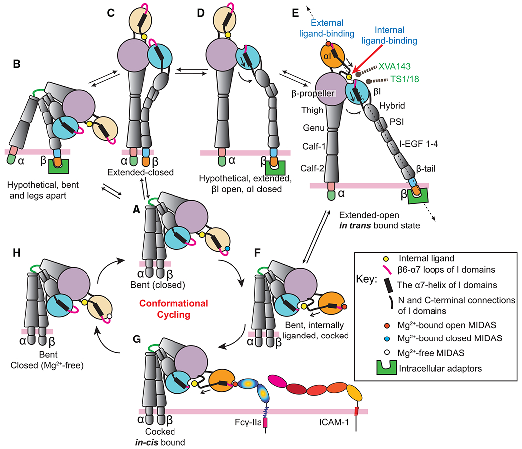
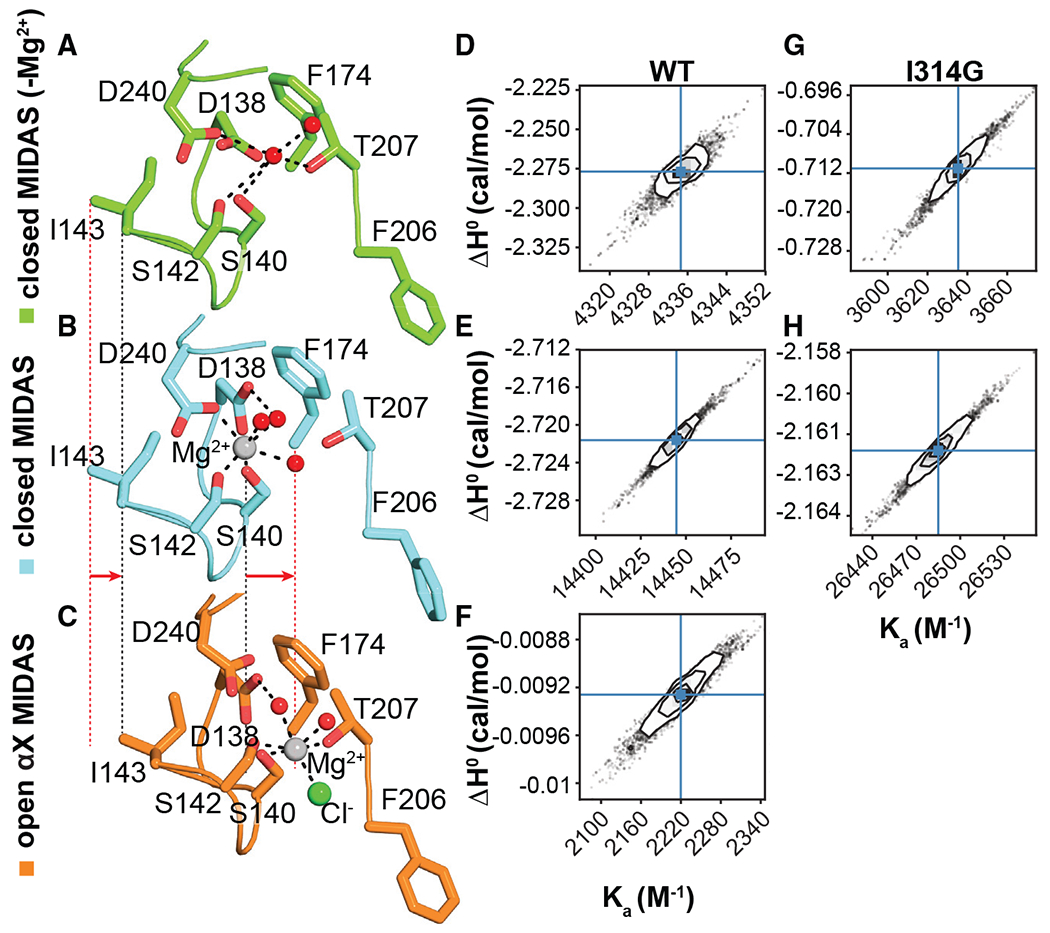
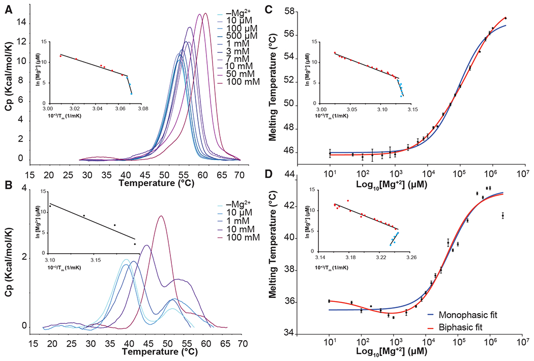
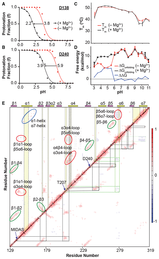
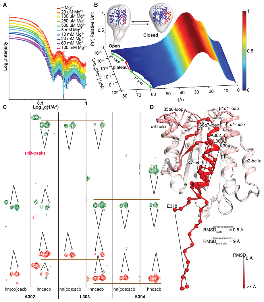
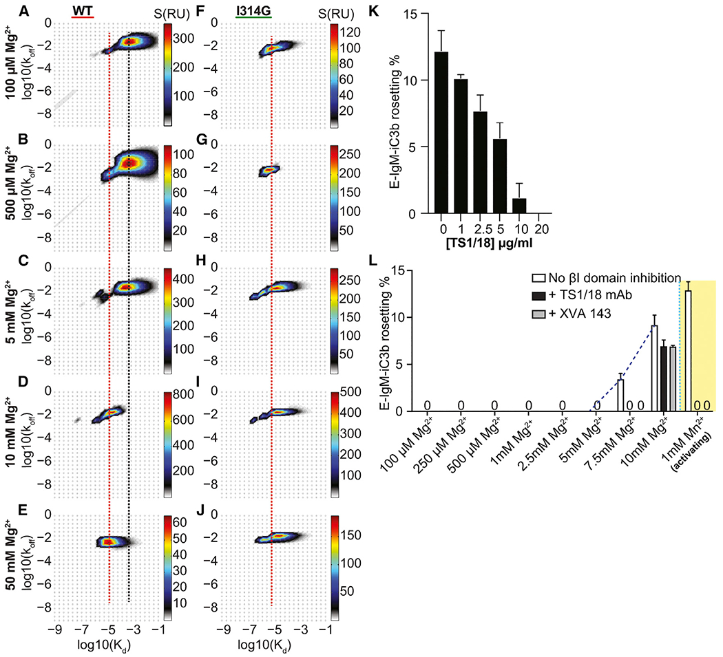
Allosteric activation and silencing of leukocyte β2-integrins transpire through cation-dependent structural changes, which mediate integrin biosynthesis and recycling, and are essential to designing leukocyte-specific drugs. Stepwise addition of Mg2+ reveals two mutually coupled events for the αXβ2 ligand-binding domain-the αX I-domain-corresponding to allostery establishment and affinity maturation. Electrostatic alterations in the Mg2+-binding site establish long-range couplings, leading to both pH- and Mg2+-occupancy-dependent biphasic stability change in the αX I-domain fold. The ligand-binding sensorgrams show composite affinity events for the αX I-domain accounting for the multiplicity of the αX I-domain conformational states existing in the solution. On cell surfaces, increasing Mg2+ concentration enhanced adhesiveness of αXβ2. This work highlights how intrinsically flexible pH- and cation-sensitive architecture endows a unique dynamic continuum to the αI-domain structure on the intact integrin, thereby revealing the importance of allostery establishment and affinity maturation in both extracellular and intracellular integrin events.

Keywords: CD11c; CD18; CP: Molecular biology; affinity maturation; allostery; integrin; αX I-domain.


Conflict of interest statement

Declaration of interests The authors declare no competing interests.

Figures

Figure 1.
Figure 1.. Schematic of the αI integrin activation mechanisms via 2 possible unique routes
Activation begins from (A) the bent/closed state and ends in (E) the extended open state. These conformational states have been observed in crystal structures, SAXS, electron microscopy, cell-based studies, or are otherwise noted as hypothetical. Route I: The bent/closed state (A) could have leg separated via the cytoplasmic adaptor binding (B) or ectodomain extension (C). The “bent and legs apart” state (B) is unlikely to exist or must be a very short-lived state. Headpiece opening and leg separation (D) would prepare the in trans external-ligand binding (E). Route II: In the second activation mechanism, the conformational cycling occurs starting in the (A) bent closed state and then progressing to the (F) cocked (Sen et al., 2013), (G) cocked-in cis bound (Saggu et al., 2018), and (H) the bent/closed-Mg2+-free states sequentially. Locations of XVA143 and TS1/18 mAb bindings are labeled, and external ligands, in trans or ICAM-1 and Fcγ-IIa in cis interactions, are noted. The Mg2+-free or Mg2+-bound MIDAS in the closed and open αI domain, when needed, were noted as white, blue, or red spheres, respectively. The β2-tail of the states that are available to intracellular interactions (B, D, and E) are shown to couple cytoplasmic adaptors.
Figure 2.
Figure 2.. Dication interactions of the αX I-domain
The MIDAS assembly in the (A) Mg2+-free closed (PDB: 1N3Y), (B) Mg2+-bound closed (PDB: 5ES4), and (C) Mg2+-bound open (PDB: 4NEH) states. Mg2+, water, and CI are shown as silver, red, and green spheres, respectively. The dashed line shows the lateral movements of Mg2+ and I143. Interactions of (D and G) Mg2+, (E and H) Mn2+, and (F) Ca2+ with the WT αX I-domain and the I314G were probed using corner plots, showing correlations between the posterior distribution for 2 energetic parameters of the binding constant(Ka) and enthalpy (ΔH0binding).
Figure 3.
Figure 3.. Effect of Mg2+ on the thermal stability of the αX I-domain
(A and B) DSC thermograms for the (A) WT αX I-domain and (B) I314G in increasing concentration of Mg2+. (C and D) Tm change in response to Mg2+concentration from DSF denaturation for the (C) WT αX I-domain and (D) I314G were plotted and fitted to monophasic (blue) or biphasic transition (red). Plots of Van ’t Hoff linear dependence between 1/Tm and In[Mg2+] are shown as the inset for the WT αX I-domain DSC and DSF dataset and the I314G construct DSF dataset.
Figure 4.
Figure 4.. Probing the ionization states of the conserved MIDAS Asps and correlated motions of the αX I-domain fold
(A and B) Shifts in pKa of (A) D138 and (B) D240 were calculated using nonequilibrium molecular dynamics and Monte Carlo simulations for the WT αX I-domain. (C and D) Linkage analysis of pH dependence in the range of 3–11, probed by (C) the Tm change and (D) the thermodynamic stability (ΔGunfolding) of the WT αX I-domain in Mg2+-free and 1 mM Mg2+. Unfolding free energy differences shown (ΔΔGunfolding, blue line) is significant at the pH range of 3–6, and plateaus to zero at the pH values higher than 6. (E) Residue cross-correlations (RCC) calculated from the full set of normal modes of the Mg2+-bound αX I-domain Mg2+. Map is color-coded, ranging from dark blue for high anticorrelations to dark red for high correlations. Routes that provide the 2 long-range coupling from MIDAS to the β6-α7 loop are drawn as black and green dashed lines, and residues that show NMR splitting are highlighted as the vertical yellow-shaded areas.
Figure 5.
Figure 5.. Effect of Mg2+ on the αX I-domain structure
(A and B) (A) SWAXS intensity I(q) data and (B) the interpolated 3D pairwise distribution curves (P(r)) derived from the SWAXS intensity in increasing concentration of Mg2+. (C) Representative strip plots from the 3D-HNCACB and HN(CO)CACB spectra, illustrating the split peaks and connectivities of 13Cα/13Cβ chemical shifts. The pair of HNCACB and HN(CO)CACB NMR strips for 3 exemplary residues, A302, L303, and K304, are separated by a gray line, and each residue pair is separated by a black line. The brown lines that cross one pair to the next NMR strip indicate and validate chemical shift assignment for the Cα (red) and Cβ (green) resonances. (D) All of the residues having split peaks are mapped to 2 closed and open states, and the Cα atoms of those residues are shown as spheres.
Figure 6.
Figure 6.. Effect of Mg2+ on αXβ2 affinity
(A–J) Distribution of the binding kinetics of fibrinogen with (A–E) the WT αX I-domain and (F–J) I314G construct in varying Mg2+ concentrations shown as a 2D grid (Kd and Koff). The black and red lines represent and are centered on the low- and high-affinity Kd values on the 2D grid. (K and L) Inhibitory effect of TS1/18 mAb concentration on the iC3b rosetting experiment (K). Effect of the increasing concentration of Mg2+ ion on the cell surface expressed (L) human αXβ2 rosetting with the opsonized sheep erythrocytes. The yellow-shaded area shows the Mn2+-induced αXβ2 affinity increase.

Similar articles

Cited by

References

    1. Adebamowo SN, Spiegelman D, Willett WC, and Rexrode KM (2015). Association between intakes of magnesium, potassium, and calcium and risk of stroke: 2 cohorts of US women and updated meta-analyses. Am. J. Clin. Nutr 101, 1269–1277. 10.3945/ajcn.114.100354. - DOI - PMC - PubMed
    1. Ajroud K, Sugimori T, Goldmann WH, Fathallah DM, Xiong JP, and Arnaout MA (2004). Binding affinity of metal ions to the CD11b A-domain is regulated by integrin activation and ligands. J. Biol. Chem 279, 25483–25488. 10.1074/jbc.M402901200. - DOI - PubMed
    1. Azcutia V, Routledge M, Williams MR, Newton G, Frazier WA, Manica A, Croce KJ, Parkos CA, Schmider AB, Turman MV, et al. (2013). CD47 plays a critical role in T-cell recruitment by regulation of LFA-1 and VLA-4 integrin adhesive functions. Mol. Biol. Cell 24, 3358–3368. 10.1091/mbc.E13-01-0063. - DOI - PMC - PubMed
    1. Bajic G, Yatime L, Sim RB, Vorup-Jensen T, and Andersen GR (2013). Structural insight on the recognition of surface-bound opsonins by the integrin I domain of complement receptor 3. Proc. Natl. Acad. Sci. USA 110, 16426–16431. 10.1073/pnas.1311261110. - DOI - PMC - PubMed
    1. Baldwin ET, Sarver RW, Bryant GL Jr., Curry KA, Fairbanks MB, Finzel BC, Garlick RL, Heinrikson RL, Horton NC, Kelley LL, et al. (1998). Cation binding to the integrin CD11b I domain and activation model assessment. Structure 6, 923–935. 10.1016/s0969-2126(98)00093-8. - DOI - PubMed

Publication types