Review
. 2022 Sep;24(9):905-933.
doi: 10.1177/1098612X221118761.

2022 AAFP/EveryCat Feline Infectious Peritonitis Diagnosis Guidelines

Affiliations
Review

2022 AAFP/EveryCat Feline Infectious Peritonitis Diagnosis Guidelines

Vicki Thayer et al. J Feline Med Surg. 2022 Sep.

Erratum in

Abstract

Clinical importance: Feline infectious peritonitis (FIP) is one of the most important infectious diseases and causes of death in cats; young cats less than 2 years of age are especially vulnerable. FIP is caused by a feline coronavirus (FCoV). It has been estimated that around 0.3% to 1.4% of feline deaths at veterinary institutions are caused by FIP.

Scope: This document has been developed by a Task Force of experts in feline clinical medicine as the 2022 AAFP/EveryCat Feline Infectious Peritonitis Diagnosis Guidelines to provide veterinarians with essential information to aid their ability to recognize cats presenting with FIP.

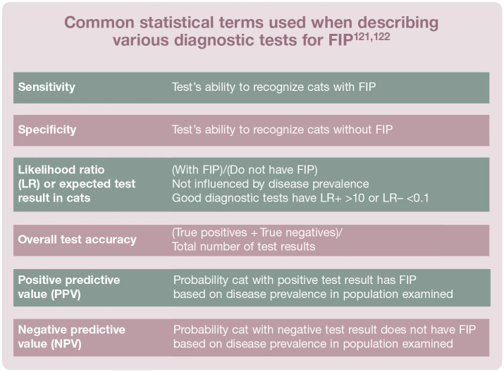
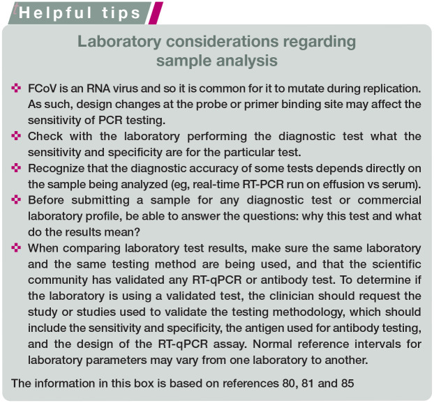
Testing and interpretation: Nearly every small animal veterinary practitioner will see cases. FIP can be challenging to diagnose owing to the lack of pathognomonic clinical signs or laboratory changes, especially when no effusion is present. A good understanding of each diagnostic test's sensitivity, specificity, predictive value, likelihood ratio and diagnostic accuracy is important when building a case for FIP. Before proceeding with any diagnostic test or commercial laboratory profile, the clinician should be able to answer the questions of 'why this test?' and 'what do the results mean?' Ultimately, the approach to diagnosing FIP must be tailored to the specific presentation of the individual cat.

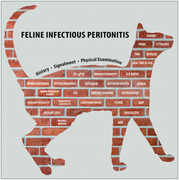
Relevance: Given that the disease is fatal when untreated, the ability to obtain a correct diagnosis is critical. The clinician must consider the individual patient's history, signalment and comprehensive physical examination findings when selecting diagnostic tests and sample types in order to build the index of suspicion 'brick by brick'. Research has demonstrated efficacy of new antivirals in FIP treatment, but these products are not legally available in many countries at this time. The Task Force encourages veterinarians to review the literature and stay informed on clinical trials and new drug approvals.

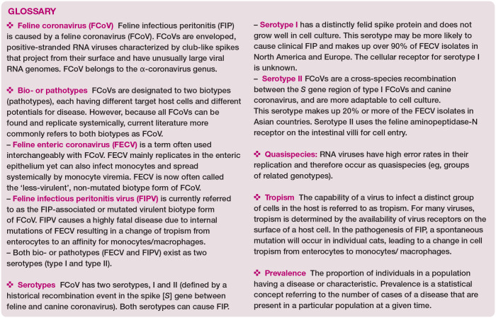
Keywords: AFAST; FCoV; FIP; Feline infectious peritonitis; PCR; PCR testing; RNA; RNA virus; Rivalta; analysis; antibody; blood test; cytology; diagnosis; effusion; feline coronavirus; fluid; imaging; infection; laboratory sample; lesion; polymerase chain reaction; screening; virus.


Conflict of interest statement

The Task Force members have no conflicts of interest to declare.

Figures

Figure 1
Figure 1
Diagnostic work-up for FIP. The veterinarian must consider the patient’s history, signalment and physical examination findings, and then select diagnostic tests and sample types based on these, in order to build the index of suspicion ‘brick by brick’. ADR = ‘ain’t doing right’. For explanation of other abbreviations, see box on page 906. This figure is available to download from the supplementary material (supplemental figure 8). Graphic designed by Vicki Thayer and Susan Gogolski, based on Melissa Kennedy’s diagnostic brick wall
Figure 2
Figure 2
Kitten from a rescue group showing lethargy (listlessness), a common early sign of FIP. LIkewise, failure to thrive or unthriftiness is a frequent complaint in a kitten or young cat with FIP. This kitten eventually developed seizures from neurological FIP. Courtesy of Julie Jacobs
Figure 3
Figure 3
Male Ragdoll with a history of anorexia, fever and weight loss of 2 lb (0.9 kg) over 2 weeks, as well as effusion, caused by FIP. Courtesy of Stacey DeVaney
Figure 4
Figure 4
Female Oriental Shorthair from a cattery, showing significant weight loss. Jaundice and effusion were also present. Courtesy of Lisa Callaway
Figure 5
Figure 5
(a) Female spayed Persian showing depression, weakness and tremors (see supplemental video 10, which shows this patient’s neurological signs). (b) Same cat with progression of neurological signs to decreased consciousness, weakness and focal seizures. Images courtesy of Tammy Evans
Figure 6
Figure 6
Cortical blindness in a male Bengal manifesting as bilateral dilated pupils. Courtesy of Glenn Olah
Figure 7
Figure 7
Five-year-old male castrated domestic shorthair cat with non-effusive FIP presenting as progressive weight loss, anemia and development of blindness. The cat was PCR positive on FNAs of kidney, liver, spleen and MLNs. Courtesy of Matthew Kornya
Figure 8
Figure 8
Oriental Shorthair with anterior uveitis, and corneal edema due to development of glaucoma. Courtesy of Lisa Callaway
Figure 9
Figure 9
Weight loss and progression of neurological signs accompanied the presence of anisocoria in this 12-year-old male neutered domestic shorthair cat. Courtesy of Glenn Olah
Figure 10
Figure 10
(a,b) Two cats demonstrating subtle differences in the clinical presentation of anterior uveitis caused by FIP: iris color changes, haziness in the anterior chamber (hypopyon or hyphema) and lesion(s) (keratic precipitates) in the cornea. The young cat in (b) has additional evidence of irregular pupils, which can occur with anterior uveitis caused by FIP. Repeated ophthalmoscopic examinations are indicated in difficult-to-diagnose FIP cases. Images courtesy of Marybeth Rymer (a) and Haley Batemen (b)
Figure 11
Figure 11
(a) Uveitis and leakage of fibrin into the anterior chamber of the eye. (b) Rubeosis iridis and a clump of fibrin. Images courtesy of Jessica Meekins
Figure 12
Figure 12
(a,b) Cat with abdominal effusion (ascites) presenting with abdominal distension, weight loss and decreased muscle mass. This patient is shown in supplemental video 11. Images courtesy of Stephanie Newton
Figure 13
Figure 13
Ventral inguinal area of an 8-year-old male neutered domestic shorthair cat with significant dermal vasculitis and neutrophilic splenitis that later developed effusion and was diagnosed with FIP. Cats with skin changes can be difficult-to-diagnose FIP cases. These images show the initial (a) and later (b) presentation of the vasculitis. Courtesy of Matthew Kornya Courtesy of Matthew Kornya
Figure 14
Figure 14
Essential components of a comprehensive physical examination. (a,b) Fever is one of the earliest non-specific signs of FIP. A temperature should be measured during routine visits in a cat friendly manner (eg, using an ear thermometer); if elevated, a rectal thermometer is used to confirm the existence of fever. (c) Palpation of the abdomen is an essential part of the physical examination for detecting enlarged MLNs, or nodular irregularities on the surface of kidneys or liver due to pyogranulomatous lesions. In addition, abdominal effusion, which is common in cats with FIP, can be noted by the presence of a fluid wave during this procedure. (d) Ocular involvement can be subtle yet creates a number of different changes including keratic precipitates, anterior and/or posterior uveitis, anisocoria and retinal detachment. A full opththalmic examination should be incorporated into the physical examination to obtain a diagnosis of FIP. Image (a) courtesy of Dianne Brown; images (b), (c), and (d) courtesy of Glenn Olah
Figure 15 (a)
Figure 15 (a)
Abdominal effusion due to FIP collected for sample analysis. (b) Initial assessment of an effusion sample due to FIP. The yellow color and sticky consistency (reflecting the high protein concentration in the fluid) is typical. Images courtesy of Emi Barker (a) and Matthew Kornya (b)
Figure 16a
Figure 16a
FIP (effusion): mixed cell population (non-degenerate neutrophils, macrophages and one lymphocyte) dispersed with a few erythrocytes in a granular eosinophilic proteinaceous background commonly observed in FIP effusions. Modified Wright stain, x 100
Figure 16b
Figure 16b
Splenic mast cell tumor: the mast cells are well differentiated with numerous purple-staining metachromatic granules that often obscure the nucleus. Splenic FNA, Wright-Giemsa stain, x 20
Figure 16c
Figure 16c
Septic peritonitis (effusion): there is an abundance of neutrophils present; some are swollen with dispersed nuclear chromatin, others have cytoplasmic vacuoles with small rod-shaped bacteria within the cytoplasm. Cytospin preparation, Wright-Giemsa stain, x 100
Figure 16d
Figure 16d
Mixed thoracic fluid (circulatory): there is a mixture of cells present, including small and intermediate sized lymphocytes, neutrophils, macrophages and red blood cells. The vacuolated macrophages exhibit erythrophagy. Cytospin preparation, Wright-Giemsa stain, x 50
Figure 16e
Figure 16e
Cryptococcosis: this is an impression smear of a mass on the neck of a cat; there are macrophages with fewer neutrophils, a plasma cell and many yeast organisms, some with the large capsule typical of Cryptococcus species. One organism (arrow) exhibits the narrow-based budding seen with this yeast. Wright-Giemsa stain, x 50
Figure 16f
Figure 16f
Normal lymph node: there is a mixed lymphoid population present in this FNA of a lymph node. Small lymphocytes are predominant but plasma cells, lymphoblasts, nuclear debris and lymphoglandular bodies are present, as well as occasional macrophages. Wright-Giemsa stain, x 50
Figure 16g
Figure 16g
Intestinal lymphoma (effusion): this is a large cell variant of lymphoma with numerous lymphoblasts; small lymphocytes and neutrophils are also present. Cytospin sediment preparation, Wright-Giemsa stain, x 100 Images courtesy of Sally Lester (a-f) and Christopher Lanier (g)
Figure 17
Figure 17
Positive Rivalta’s test. Courtesy of Katrin Hartmann, LMU Munich
Figure 18
Figure 18
Positive AFAST at the subxiphoid (Diaphragmatico-Hepatic [DH] view) demonstrating a typical anechoic accumulation of free fluid between the diaphragm and liver. By looking cranial to the diaphragm, pleural and pericardial effusion and lung pathology can also be detected
Figure 19
Figure 19
Positive AFAST (Spleno-Renal view) in the region of the costal arch and sublumbar muscles, demonstrating a typical anechoic accumulation of free fluid along the cranial pole of the left kidney and tail of the spleen. The target organs are the left kidney and spleen, although in most cats both kidneys may be seen at this view
Figure 20
Figure 20
Positive AFAST (Cysto-Colic view [a] with a urine-filled bladder) demonstrating a typical anechoic accumulation of free fluid between the urinary bladder and the body wall in the most gravity-dependent region called the Cysto-Colic Pouch. The target organ is the urinary bladder. Small intestine will also be in view
Figure 21
Figure 21
Positive AFAST (Hepato-Renal Umbilical view) at the level of the umbilicus demonstrating a typical anechoic accumulation of free fluid between loops of intestine (arrow), the body wall, and omentum in the most gravity-dependent region called the Umbilical Pouch. The target organs are the small intestine and spleen
Figure 22
Figure 22
Mesenteric lymphadenopathy. The hypoechoic structure in the mid-abdomen may be screened for during AFAST and its target organ approach and detected during complete abdominal ultrasound studies. In (a) the image is unlabeled and in (b) labeled with the mesenteric lymph node outlined with an oval and small intestine (SI). Enlargement has been defined by measurements of thickness of being no more than 5 mm; however, some may be considered normal up to 7-8 mm. The ratio of short axis to long axis is another parameter that may be used, with normal <0.5. Images courtesy of, and with permission from, Dr Stephanie Lisciandro, Hill Country Veterinary Specialists and FASTVET.com, Spicewood, Texas, USA © 2021
Figure 23
Figure 23
Renomegaly, Poor Architecture, Perirenal Fluid. In (a) and (b) there is renomegaly and a small rim of subcapsular fluid that may be suspected during AFAST. The length should never exceed 4.5 cm in a cat and this kidney clearly measures over 6 cm. Measurements are also increased for width and height in its transverse plane. In (c) and (d) there is obvious loss of expected architecture in both kidneys; again, a finding that could be suspected during AFAST using its target organ approach. There is perirenal fluid associated with these kidneys as well. In (e) is another example of perirenal fluid. By detecting these abnormalities during AFAST as part of the initial diagnostics of the cat, the information is captured quickly and the diagnostic plan is potentially more streamlined. LK = left kidney. Images courtesy of, and with permission from, Drs Gregory and Stephanie Lisciandro, Hill Country Veterinary Specialists and FASTVET.com, Spicewood, Texas, USA; and Point-of-Care Ultrasound Techniques for the Small Animal Practitioner, 2nd edition © 2021
Figure 24
Figure 24
Right lateral (a) and ventrodorsal (b) thoracic radiographs of a cat with pleural effusion. Images courtesy of, and with permission from, Dr Gregory Lisciandro, Hill Country Veterinary Specialists and FASTVET.com, Spicewood, Texas, USA; and Point-of-Care Ultrasound Techniques for the Small Animal Practitioner, 2nd edition © 2021134
Figure 25
Figure 25
Pleural effusion located in the cardiac diaphragmatic pouch. During the ‘TFAST slide’, the probe is moved caudally from the heart to the diaphragm where the most gravity-dependent area is located, the cardiac diaphragmatic pouch. Images courtesy of, and with permission from, Dr Gregory Lisciandro, Hill Country Veterinary Specialists and FASTVET.com, Spicewood, Texas, USA; and Point-of-Care Ultrasound Techniques for the Small Animal Practitioner, 2nd edition © 2021
Figure 26
Figure 26
(a,b,c) Pleural effusion located in the cardiac cervical pouch. During the ‘TFAST slide’, the probe is moved cranially across the heart to the cardiac cervical pouch, which is the most gravity-dependent area in the standing or sternal patient positions and is located cranial to the heart, which is identified by the arrows. Images courtesy of, and with permission from, Dr Gregory Lisciandro, Hill Country Veterinary Specialists and FASTVET.com, Spicewood, Texas, USA; and Point-of-Care Ultrasound Techniques for the Small Animal Practitioner, 2nd edition © 2021
Figure 27
Figure 27
Pleural and pericardial effusion. The entire heart is visible. Images courtesy of, and with permission from, Dr Gregory Lisciandro, Hill Country Veterinary Specialists and FASTVET.com, Spicewood, Texas, USA; and Point-of-Care Ultrasound Techniques for the Small Animal Practitioner, 2nd edition © 2021
None
None
None
None
None
None
None
None
None
None
None
None
None
None
None
None

Similar articles

Cited by

References

    1. Holzworth J. Some important disorders of cats. Cornell Vet 1963; 53: 157–160. - PubMed
    1. Klein-Richers U, Hartmann K, Hofmann-Lehmann R, et al.. Prevalence of feline coronavirus shedding in German catteries and associated risk factors. Viruses 2020; 12: 1000–1013. - PMC - PubMed
    1. Riemer F, Kuehner KA, Ritz S, et al.. Clinical and laboratory features of cats with feline infectious peritonitis – a retrospective study of 231 confirmed cases (2000-2010). J Feline Med Surg 2016; 18: 348–356. - PMC - PubMed
    1. Pesteanu-Somogyi LD, Radzai C, Pressler BM. Prevalence of feline infectious peritonitis in specific cat breeds. J Feline Med Surg 2006; 8: 1–5. - PMC - PubMed
    1. Rohrbach BW, Legendre AM, Baldwin CA, et al.. Epidemiology of feline infectious peritonitis among cats examined at veterinary medical teaching hospitals. J Am Vet Med Assoc 2001; 218: 1111–1115. - PubMed

Publication types

LinkOut - more resources