. 2022 Oct 1;323(4):E389-E401.
doi: 10.1152/ajpendo.00078.2022. Epub 2022 Aug 24.

Glucagon receptor signaling at white adipose tissue does not regulate lipolysis

Affiliations

Glucagon receptor signaling at white adipose tissue does not regulate lipolysis

Anastasiia Vasileva et al. Am J Physiol Endocrinol Metab. .

Abstract

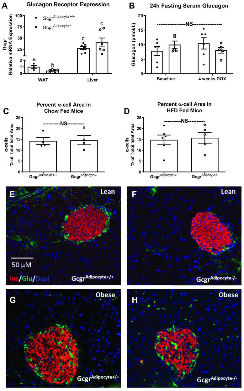
Although the physiological role of glucagon receptor signaling in the liver is well defined, the impact of glucagon receptor (Gcgr) signaling on white adipose tissue (WAT) continues to be debated. Although numerous studies propose that glucagon stimulates WAT lipolysis, we lack evidence that physiological concentrations of glucagon regulate WAT lipolysis. In turn, we performed studies in both wild-type and WAT Gcgr knockout mice to determine if glucagon regulates lipolysis at WAT in the mouse. We assessed the effects of fasting and acute exogenous glucagon administration in wild-type C57BL/6J and GcgrAdipocyte+/+ versus GcgrAdipocyte-/- mice. Using an ex vivo lipolysis protocol, we further examined the direct effects of glucagon on physiologically (fasted) and pharmacologically stimulated lipolysis. We found that adipocyte Gcgr expression did not affect fasting-induced lipolysis or hepatic lipid accumulation in lean or diet-induced obese (DIO) mice. Acute glucagon administration did not affect serum nonesterified fatty acids (NEFA), leptin, or adiponectin concentration, but did increase serum glucose and FGF21, regardless of genotype. Glucagon did not affect ex vivo lipolysis in explants from either GcgrAdipocyte+/+ or GcgrAdipocyte-/- mice. Gcgr expression did not affect fasting-induced or isoproterenol-stimulated lipolysis from WAT explants. Moreover, glucagon receptor signaling at WAT did not affect body weight or glucose homeostasis in lean or DIO mice. Our studies have established that physiological levels of glucagon do not regulate WAT lipolysis, either directly or indirectly. Given that glucagon receptor agonism can improve dyslipidemia and decrease hepatic lipid accumulation, it is critical to understand the tissue-specific effects of glucagon receptor action. Unlike the crucial role of hepatic glucagon receptor signaling in maintaining glucose and lipid homeostasis, we observed no metabolic consequence of WAT glucagon receptor deletion.NEW & NOTEWORTHY It has been postulated that glucagon stimulates lipolysis and fatty acid release from white adipose tissue. We observed no metabolic effects of eliminating or activating glucagon receptor signaling at white adipose tissue.

Keywords: fasting; glucagon; lipolysis; type 2 diabetes; white adipose tissue.


Conflict of interest statement

No conflicts of interest, financial or otherwise, are declared by the authors.

Figures

None
Graphical abstract
Figure 1.
Figure 1.
Acute intraperitoneal glucagon administration in wild-type mice. Serum glucagon, glucose, insulin, and NEFA concentrations in wild-type C57BL/6J mice fasted for 4 (A, C, E, and G) or 16 (B, D, F, and H) h (n = 5–7 mice/group for all except 4-h fasted insulin, n = 3–5/group). Mice were injected with saline (time 0) or glucagon (5 µg/kg) and euthanized at 15, 30, or 60 min after intraperitoneal injection. Data presented as means ± SE. a,bSuperscript letters that differ indicate differences within group, P < 0.05. One-way ANOVA with Tukey’s adjustment for multiple comparisons. NEFA, nonesterified fatty acids; NS, not significant.
Figure 2.
Figure 2.
Ex vivo lipolysis from gonadal adipose tissue explants. Explant NEFA release in response to bath application of isoproterenol, forskolin and glucagon was assessed in 4-h and 16-h fasted mice. Isoproterenol and forskolin stimulated explant NEFA release from wild-type C57BL/6J mice fasted for 4 h (A; n = 10 mice) or 16 h (B; n = 8). Media NEFA concentrations induced by glucagon in 4 h (C; n = 10) and 16 h (D; n = 8) fasted wild-type C57BL/6J mice. NEFA release from explants collected from GcgrAdipocyte+/+ vs. GcgrAdipocyte−/− mice fasted for 4 h (n = 4) or 16 h (Gcgradipocyte+/+: n = 7, Gcgradipocyte−/−: n = 10) and treated with forskolin (E and F) or glucagon (G and H). All studies were performed in triplicate explants from each mouse. Data presented as means ± SE; paired samples t test. NEFA, nonesterified fatty acids; NS, not significant.
Figure 3.
Figure 3.
Gcgradipocyte−/− mice fed a low-fat diet have normal fasting-induced changes in serum NEFA concentration and hepatic lipid accumulation. Serum NEFA (A) and hepatic triglyceride (B) concentrations in mice fasted for 4 h (n = 6 or 7 Gcgradipocyte+/+ and n = 4–6 Gcgradipocyte−/− mice) and 16 h (n = 7 Gcgradipocyte+/+ and n = 10–11 Gcgradipocyte−/−). Serum NEFA (C) and glucose (D) concentrations in 16-h fasted Gcgradipocyte+/+ and Gcgradipocyte−/− mice injected with saline and glucagon (n = 6/genotype). a,bSuperscript letters that differ indicate differences within group, P < 0.01. Data presented as means ± SE. A and B: two-way ANOVA with Tukey’s adjustment for multiple comparisons. C and D: paired samples t test (cross over glucagon responsivity test). NEFA, nonesterified fatty acids; NS, not significant.
Figure 4.
Figure 4.
Gcgradipocyte−/− mice fed a low fat diet have a normal response to oral glucose. Body weight (A), oral glucose tolerance (OGTT; B), OGTT area under the curve (C), glucose-stimulated insulin (D), insulin tolerance test (ITT; E), and ITT area under the curve in lean mice (F). For body weight, n = 11 Gcgradipocyte+/+ and n = 13 Gcgradipocyte−/− mice. For OGTT and glucose-stimulated insulin, n = 13 Gcgradipocyte+/+ and n = 7 Gcgradipocyte−/− mice. For ITT, n = 6 Gcgradipocyte+/+ and n = 6 Gcgradipocyte−/− mice). a,bSuperscript letters that differ indicate differences within group, P < 0.01; two-way ANOVA with Tukey’s adjustment for multiple comparisons for (A) body weight and (D) glucose stimulated insulin, independent t test for (C) OGTT and (F); ITT AUC. Data presented as means ± SE. AUC, area under the curve; NS, not significant.
Figure 5.
Figure 5.
Gcgr gene expression, fasting glucagon, and α-cell abundance in Gcgradipocyte+/+ and Gcgradipocyte−/− mice. A: relative mRNA expression of glucagon receptor in liver (n = 7 mice/genotype) and gonadal adipose tissue (n = 4 Gcgradipocyte+/+ and n = 7 Gcgradipocyte−/−) in lean mice. B: 24-h fasting serum glucagon in lean Gcgradipocyte+/+ (n = 6) and Gcgradipocyte−/− (n = 5) mice before and after 4 wk of doxycycline induction. α-Cell percentage of total islet area in (C) lean (n = 4/genotype) and (D) obese (n = 6 Gcgradipocyte+/+ and n = 5 Gcgradipocyte−/−) mice with representative photos (EH). a,b,cSuperscript letters that differ indicate differences within group, P < 0.05; two-way ANOVA with Tukey’s adjustment for multiple comparisons for (A) mRNA expression of glucagon receptor and (B) fasting serum glucagon, independent t test for (C and D) α-cell percentage of total islet area. Data presented as means ± SE. NS, not significant.

Similar articles

Cited by

References

    1. Longuet C, Robledo AM, Dean ED, Dai C, Ali S, McGuinness I, de Chavez V, Vuguin PM, Charron MJ, Powers AC, Drucker DJ. Liver-specific disruption of the murine glucagon receptor produces α-cell hyperplasia: evidence for a circulating α-cell growth factor. Diabetes 62: 1196–1205, 2013. doi:10.2337/db11-1605. - DOI - PMC - PubMed
    1. Zeigerer A, Sekar R, Kleinert M, Nason S, Habegger KM, Müller TD. Glucagon’s metabolic action in health and disease. Compr Physiol 11: 1759–1783, 2021. doi:10.1002/cphy.c200013. - DOI - PMC - PubMed
    1. Galsgaard KD, Pedersen J, Knop FK, Holst JJ, Wewer Albrechtsen NJ. Glucagon receptor signaling and lipid metabolism. Front Physiol 10: 413, 2019. doi:10.3389/fphys.2019.00413. - DOI - PMC - PubMed
    1. Longuet C, Sinclair EM, Maida A, Baggio LL, Maziarz M, Charron MJ, Drucker DJ. The glucagon receptor is required for the adaptive metabolic response to fasting. Cell Metab 8: 359–371, 2008. doi:10.1016/j.cmet.2008.09.008. - DOI - PMC - PubMed
    1. Nason SR, Kim T, Antipenko JP, Finan B, DiMarchi R, Hunter CS, Habegger KM. Glucagon-receptor signaling reverses hepatic steatosis independent of leptin receptor expression. Endocrinology 161: bqz013, 2020. doi:10.1210/endocr/bqz013. - DOI - PMC - PubMed

Publication types

LinkOut - more resources