. 2022 Sep 14;42(37):7077-7093.
doi: 10.1523/JNEUROSCI.2263-21.2022. Epub 2022 Aug 24.

Adult Born Dentate Granule Cell Mediated Upregulation of Feedback Inhibition in a Mouse Model of Traumatic Brain Injury

Affiliations

Adult Born Dentate Granule Cell Mediated Upregulation of Feedback Inhibition in a Mouse Model of Traumatic Brain Injury

Young-Jin Kang et al. J Neurosci. .

Abstract

Post-traumatic epilepsy (PTE) and behavioral comorbidities frequently develop after traumatic brain injury (TBI). Aberrant neurogenesis of dentate granule cells (DGCs) after TBI may contribute to the synaptic reorganization that occurs in PTE, but how neurogenesis at different times relative to the injury contributes to feedback inhibition and recurrent excitation in the dentate gyrus is unknown. Thus, we examined whether DGCs born at different postnatal ages differentially participate in feedback inhibition and recurrent excitation in the dentate gyrus using the controlled cortical impact (CCI) model of TBI. Both sexes of transgenic mice expressing channelrhodopsin2 (ChR2) in postnatally born DGCs were used for optogenetic activation of three DGC cohorts: postnatally early born DGCs, or those born just before or after CCI. We performed whole-cell patch-clamp recordings from ChR2-negative, mature DGCs and parvalbumin-expressing basket cells (PVBCs) in hippocampal slices to determine whether optogenetic activation of postnatally born DGCs increases feedback inhibition and/or recurrent excitation in mice 8-10 weeks after CCI and whether PVBCs are targets of ChR2-positive DGCs. In the dentate gyrus ipsilateral to CCI, activation of ChR2-expressing DGCs born before CCI produced increased feedback inhibition in ChR2-negative DGCs and increased excitation in PVBCs compared with those from sham controls. This upregulated feedback inhibition was less prominent in DGCs born early in life or after CCI. Surprisingly, ChR2-positive DGC activation rarely evoked recurrent excitation in mature DGCs from any cohort. These results support that DGC birth date-related increased feedback inhibition in of DGCs may contribute to altered excitability after TBI.SIGNIFICANCE STATEMENT Dentate granule cells (DGCs) control excitability of the dentate gyrus through synaptic interactions with inhibitory GABAergic interneurons. Persistent changes in DGC synaptic connectivity develop after traumatic brain injury, contributing to hyperexcitability in post-traumatic epilepsy (PTE). However, the impact of DGC neurogenesis on synaptic reorganization, especially on inhibitory circuits, after brain injury is not adequately described. Here, upregulation of feedback inhibition in mature DGCs from male and female mice was associated with increased excitation of parvalbumin-expressing basket cells by postnatally born DGCs, providing novel insights into underlying mechanisms of altered excitability after brain injury. A better understanding of these inhibitory circuit changes can help formulate hypotheses for development of novel, evidence-based treatments for post-traumatic epilepsy by targeting birth date-specific subsets of DGCs.

Keywords: adult neurogenesis; mossy fiber sprouting; optogenetics; parvalbumin-expressing basket cell; patch-clamp; post-traumatic epilepsy.


Figures

Figure 1.
Figure 1.
Experimental design of birth date-based expression of ChR2/EYFP in early born and adult born DGCs and light-evoked APs in a ChR2-positive DGC. A, To label early born or adult born DGCs with ChR2, Gli1-CreERT2::ChR2 double-transgenic mice were produced and injected with tamoxifen (i.p.) at one of three periods, postnatal 8-12 d, postnatal 5-7 weeks, and postnatal 7-9 weeks. All mice underwent either CCI injury or craniotomy (sham control) at 7 weeks of age and were killed 8-10 weeks after surgery for electrophysiological and immunohistochemical experiments. B, Dentate gyri ipsilateral to sham surgery expressing ChR2/EYFP in early born or adult born (before and after surgery) DGCs after injecting tamoxifen during one of three periods shown in A. Green and blue represent EYFP and DAPI staining, respectively. C, C′, ChR2/EYFP-expressing adult born DGC recovered after filling with biocytin during a patch clamp recording, as in D and E. Biocytin-filled cell DGC morphology (asterisk indicates soma; arrowheads indicate dendrites; arrow indicates axon) was analyzed post hoc. C, Biocytin-filled DGC (red). C′, The same DGC was EYFP labeled. D, Firing properties of a DGC from a sham control mouse expressing ChR2/EYFP. The DGC was held at resting membrane potential (–82 mV) before it produced accommodating AP discharges in response to 1 s depolarizing current steps (100 and 200 pA). E, Light-evoked APs in the DGC shown in D. Thirty millisecond light pulses delivered every 30 s through the 40× objective reliably produced APs in the DGC; the number of evoked APs was similar between DGCs from sham and CCI injured mice. Blue lines indicate when the blue light was delivered.
Figure 2.
Figure 2.
Aberrant mossy fiber sprouting in the inner molecular layer after severe CCI injury. TIMM and Nissl-counterstained images near injury epicenter 8-10 weeks after surgery. Dentate gyri ipsilateral to sham control (Aa) and contralateral to CCI (Cc) did not develop structural damages or mossy fiber sprouting in the inner molecular layer. Ipsilateral to CCI, cortical cavitation and hippocampal distortion were observed (B) as was intense mossy fiber sprouting in the inner molecular layer (Bb) near the injury site. Bb, Arrows indicate punctate mossy fibers in the inner molecular layer.
Figure 3.
Figure 3.
The proportion of ChR2/EYFP-expressing DGCs was not greater in the dentate gyrus of CCI mice compared with those of sham control mice. A–C, The dentate GCLs of early born (A) and adult born DGC cohorts (B, C, for pre- and post-CCI, respectively) were labeled for nuclei (blue), EYFP (green), and calbindin (red). D–F, Quantification of EYFP/calbindin double-positive DGCs. The number of EYFP/calbindin double-positive DGCs was normalized by the number of calbindin-positive nuclei within GCL. A trend of reductions after CCI was observed for early born EYFP-expressing DGCs in the dentate gyrus ipsilateral and contralateral to CCI injury compared with those of sham controls (A,D; one-way ANOVA, p = 0.052). Proportions of EYFP-expressing DGCs born around the time of injury were not statistically different (B,C,E,F). Number in each bar graph indicates the number of mice used for the study. Data are mean ± SEM.
Figure 4.
Figure 4.
Activation of DGCs born just before CCI, compared with DGCs born after CCI and early born DGCs, produces prominent synaptic inhibition of adult DGCs. A–C, Examples of responses in mature DGCs to optogenetic stimulation of adult born DGCs illustrating response variability in cells from different mice. Single light pulses (blue lines, 30 ms duration every 30 s) were used to induce APs in adult born DGCs, resulting in eIPSCs in recordings from ChR2-negative, mature DGCs. Five consecutive current traces are shown for three DGCs ipsilateral to CCI (A1–A3), CCI contralateral (B), and two DGCs from sham controls (C1,C2) after optogenetic stimulation of DGCs born just before CCI or sham surgery; all sets of traces from different mice. Inset, Expanded time scale of the top trace in A1 (dashed box) displays two eIPSC peaks. D–O, Summary of the proportion of DGCs showing eIPSCs, number of eIPSCs, eIPSC amplitude, and fold increase in feedback inhibition strength for DGCs born just before CCI (D–G), DGCs born just after CCI (H–K), and early born DGCs (L–O). The numbers above the bars indicate number of mature DGCs showing eIPSCs/number of tested mature DGCs (D,H,L) or number of mature DGCs included in eIPSC amplitude measurement (F,J,N). The numbers next to solid circles indicate number of mature DGCs included in IPSC frequency measurement (E,I,M). The relative strength of increase in feedback inhibition, based on the data from Figures 3 and 4, is shown in G, K, O, where the bar graphs represent eIPSC connectivity strength as a function of the mean mature EYFP-expressing DGC number for each cohort and normalized to that of sham controls. Data are mean ± SEM. **p < 0.01; ***p < 0.001; ipsilateral or contralateral to CCI versus sham controls. #p < 0.05; ##p < 0.01; ###p < 0.001; ipsilateral versus contralateral to CCI.
Figure 5.
Figure 5.
Excitation of GABAergic interneurons by ChR2-positive DGCs is necessary for eIPSCs. A, C, Current traces represent eIPSCs in mature DGCs before and during bath application of a GABAA receptor antagonist, SR95531 (10 μm), or a non-NMDA receptor antagonist, DNQX (20 μm). B, D, Summary of the changes in eIPSC amplitude by GABAA receptor antagonist and non-NMDA receptor antagonist. SR95531 and bicuculline (30 μm) were used in 3 and 4 mature DGCs, respectively, and the results from these 7 DGCs were combined. Replicates are DGC number. Open circles and black solid circles represent the results from individual cells and averages, respectively. Data are mean ± SEM. *p < 0.05. ***p < 0.001.
Figure 6.
Figure 6.
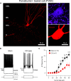
CCI-injured mice show reduced firing in PVBCs in the dentate gyrus. A, A representative image of a PVBC. The biocytin-filled PVBC was imaged using confocal microscope. Axon terminals were localized in the GCL of the dentate gyrus, whereas dendrites extended into the inner molecular layer (IML), outer molecular layer (OML), and hilus. The biocytin-labeled PVBC (red) also expressed PV (purple). B, AP firing properties of PVBCs. Evoked APs and voltage responses in PVBCs from CCI and sham control mice during 1 s depolarizing (400 pA) and hyperpolarizing (−200 pA) current steps. There is reduced AP firing in the PVBC from a CCI-injured mouse. C, The summary of AP firing frequency of PVBCs from CCI and sham control mice. n indicates number of PVBCs. Data are mean ± SEM. *p < 0.05. **p < 0.01. ***p < 0.001.
Figure 7.
Figure 7.
Activation of DGCs born just before CCI produces greater evoked EPSCs in PVBCs in the dentate gyrus from CCI-injured mice. A, Light-evoked EPSCs in PVBCs in the dentate gyrus from CCI-injured and sham control mice (blue lines, 30 ms duration, 30 s intervals). B, The summary of peak amplitudes of evoked EPSCs and (C) charge transfer. D, eEPSCs in PVBCs in the dentate gyrus from CCI and sham control mice by theta-frequency light stimuli (10 Hz, 10 stimuli, 30 ms duration). E, The summary of peak amplitude as a percent of the first peak amplitude during 10 Hz light stimulations. Data were normalized to first current amplitude in each PVBC. An increase in paired pulse ratio of evoked currents was observed in CCI injured mice versus sham controls. n indicates number of PVBCs. Data are mean ± SEM. *p < 0.05. ***p < 0.001.
Figure 8.
Figure 8.
Activation of the DGCs born just before CCI produces APs in a greater portion of PVBCs in the dentate gyrus after CCI injury. A, Similar light stimuli (30 ms duration, 30 s intervals) produced small depolarization and AP firing in PVBCs in the dentate gyrus from sham control and CCI mice, respectively. Representative 5 light-evoked voltage responses were shown for CCI-injured and sham control mice. B, The summary of proportion of PVBCs showing evoked APs. The numbers (n) above the bars indicate number of PVBCs showing evoked APs/number of tested PVBCs. *p < 0.05.
Figure 9.
Figure 9.
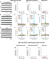
Neither adult born DGCs nor early born DGCs contribute to recurrent excitation in the dentate gyrus in CCI-injured mice. A–C, Representative examples of current traces indicate no eEPSCs in ChR2-negative mature DGCs. Single light pulses (blue lines, 30 ms duration, every 30 s) were used to produce eEPSCs. Five consecutive current traces are shown for CCI ipsilateral, CCI contralateral, and sham controls of DGCs born just before CCI. D–F, DGCs born just before CCI. G–I, DGCs born just after CCI. J–L, Early born DGCs. Summary of proportion of DGCs showing eEPSCs (D,G,J), EPSC number (E,H,K), and EPSC amplitude (F,I,L). M–O, Summary of proportion of DGCs showing eEPSCs observed in Mg2+-free ACSF containing bicuculline (30 μm). The numbers above the bars indicate number of mature DGCs showing eEPSCs/number of tested mature DGCs. The numbers next to solid circles indicate number of mature DGCs included in EPSC frequency and amplitude measurements. Data are mean ± SEM.

Comment in

Similar articles

Cited by

References

    1. Ábrahám H, Molnár JE, Sóki N, Gyimesi C, Horváth Z, Janszky J, Dóczi T, Seress L (2020) Etiology-related degree of sprouting of parvalbumin-immunoreactive axons in the human dentate gyrus in temporal lobe epilepsy. Neuroscience 448:55–70. 10.1016/j.neuroscience.2020.09.018 - DOI - PubMed
    1. Ahn S, Joyner AL (2005) In vivo analysis of quiescent adult neural stem cells responding to Sonic hedgehog. Nature 437:894–897. 10.1038/nature03994 - DOI - PubMed
    1. Althaus AL, Moore SJ, Zhang H, Du X, Murphy GG, Parent JM (2019) Altered synaptic drive onto birth dated dentate granule cells in experimental temporal lobe epilepsy. J Neurosci 39:7604–7614. 10.1523/JNEUROSCI.0654-18.2019 - DOI - PMC - PubMed
    1. Altman J, Bayer SA (1990) Migration and distribution of two populations of hippocampal granule cell precursors during the perinatal and postnatal periods. J Comp Neurol 301:365–381. 10.1002/cne.903010304 - DOI - PubMed
    1. Annegers JF, Hauser WA, Coan SP, Rocca WA (1998) A population-based study of seizures after traumatic brain injuries. N Engl J Med 338:20–24. 10.1056/NEJM199801013380104 - DOI - PubMed

Publication types