. 2022 Aug 24;13(1):4958.
doi: 10.1038/s41467-022-32665-7.

Omicron SARS-CoV-2 mutations stabilize spike up-RBD conformation and lead to a non-RBM-binding monoclonal antibody escape

Affiliations

Omicron SARS-CoV-2 mutations stabilize spike up-RBD conformation and lead to a non-RBM-binding monoclonal antibody escape

Zhennan Zhao et al. Nat Commun. .

Abstract

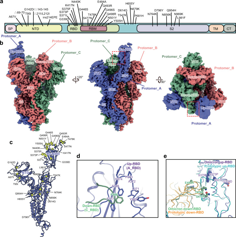
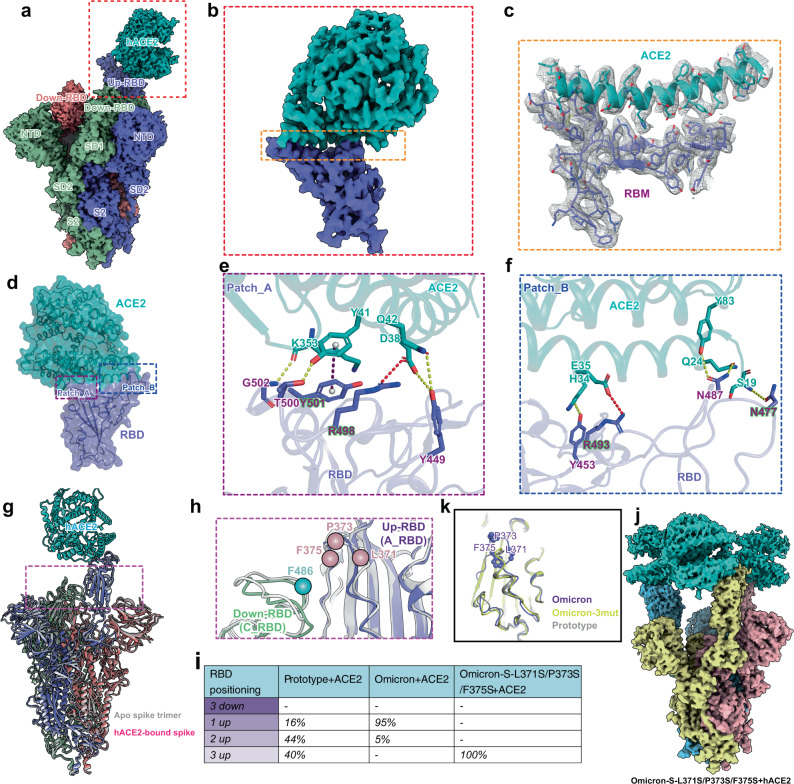
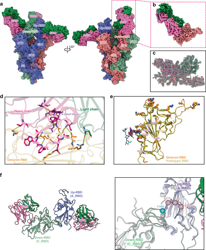
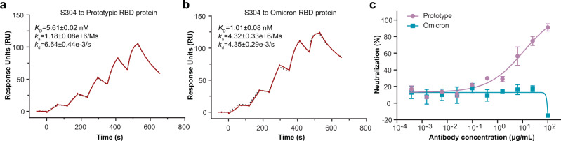
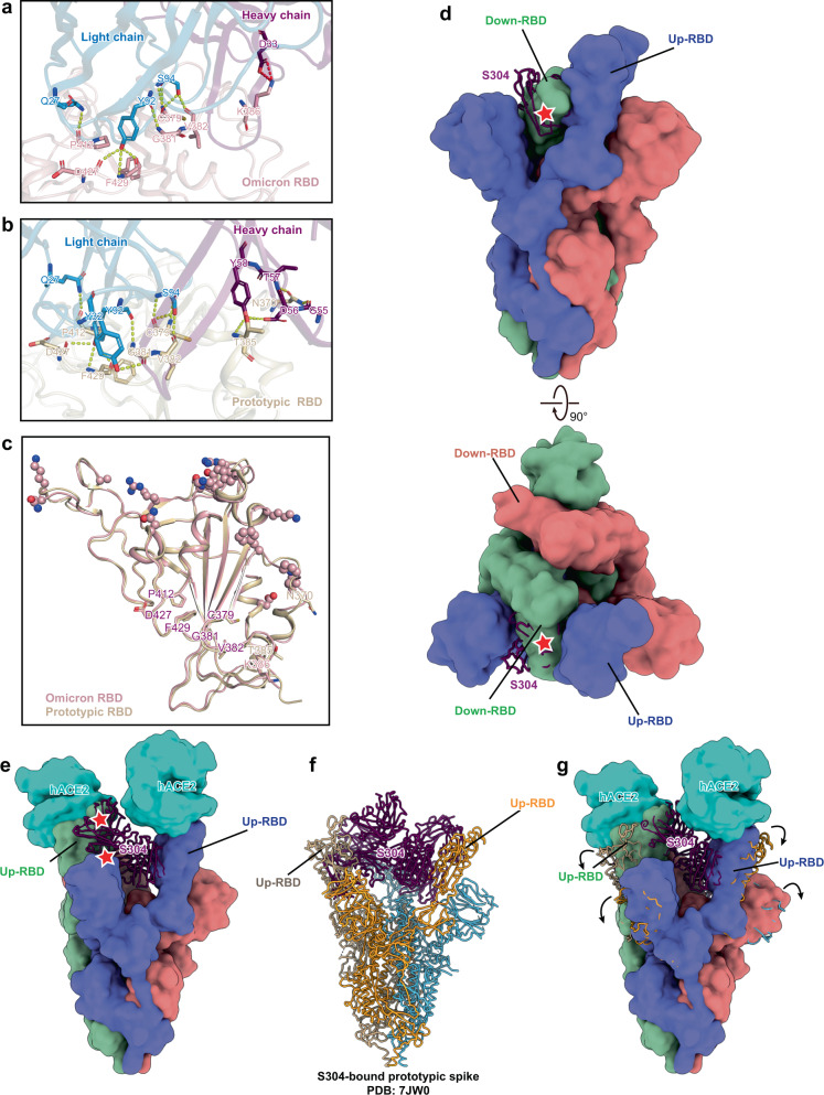
Omicron SARS-CoV-2 is rapidly spreading worldwide. To delineate the impact of emerging mutations on spike's properties, we performed systematic structural analyses on apo Omicron spike and its complexes with human ACE2 or S309 neutralizing antibody (NAb) by cryo-EM. The Omicron spike preferentially adopts the one-RBD-up conformation both before and after ACE2 binding, which is in sharp contrast to the orchestrated conformational changes to create more up-RBDs upon ACE2 binding as observed in the prototype and other four variants of concern (VOCs). Furthermore, we found that S371L, S373P and S375F substitutions enhance the stability of the one-RBD-up conformation to prevent exposing more up-RBDs triggered by ACE2 binding. The increased stability of the one-RBD-up conformation restricts the accessibility of S304 NAb, which targets a cryptic epitope in the closed conformation, thus facilitating the immune evasion by Omicron. These results expand our understanding of Omicron spike's conformation, receptor binding and antibody evasion mechanism.


Conflict of interest statement

The authors declare no competing interests.

Figures

Fig. 1
Fig. 1. Cryo-EM structure of the Omicron spike ectodomain trimer.
a A schematic diagram showing the amino acid mutations of the spike protein. b Cryo-EM map of the Omicron spike protein. Three protomers are colored in medium slate blue, light coral and dark sea green, respectively. The contact interface between down-and up-RBDs is boxed by the red dash line c Atomic structure of the Protomer_A of the Omicron spike with mutations shown as yellow spheres. d Contact interface of the Omicron up-RBD (Protomer_A, A_RBD) and neighboring down-RBD (Protomer_C, C_RBD). e Conformational comparison of the interface between A_RBD and C_RBD of the prototype (PDB: 6ZGG) and Omicron. Mutations of Omicron are shown as spheres.
Fig. 2
Fig. 2. The architecture of the ACE2-bound spike trimer of Omicron.
a Cryo-EM map of the one-RBD-up conformation of the Omicron spike protein in complex with hACE2 at 2.40 Å resolution. The ACE2 receptor is colored in cyan, and three protomers of the spike are colored corresponding to Fig. 1b. The inset box corresponds to the RBD-hACE2 region used for local refinement. b Density map of the Omicron RBD-hACE2 region at 3.24 Å resolution after local refinement for the boxed region in a. c Density map of the Omicron RBD-hACE2 interface with the fitted atomic model. Residues are shown as sticks, and density is represented in mesh. df Detailed interactions between RBD and hACE2. Yellow, red, and purple dashed lines represent hydrogen bonds, salt bridges and π-π interactions, respectively. g Superimposition of the apo and ACE2-bound spike trimers of Omicron that are reconstructed from the density maps at 3.03 Å and 2.85 Å resolution, respectively. h Conformational comparison of the interface between A_RBD and C_RBD in the apo and ACE2-bound spike trimers of Omicron. The apo spike trimer is colored in gray, and the C_RBD and A_RBD of the ACE2-bound spike trimer are colored in dark sea green and medium slate blue, respectively. The locations of residues F486 of the C_RBD, L371, P373 and F375 of the A_RBD are labeled with balls. i The relative population of RBD states observed in cryo-EM structures of ACE2-bound spikes for the prototype, Omicron and Omicron-S-L371S/P373S/F375S mutant. j Cryo-EM map of the three-RBD-up conformation of the Omicron-L371S/P373S/F375S mutant in complex with hACE2 at 3.20 Å resolution. The ACE2 receptor is colored in cyan, and three protomers of the spike trimer are colored in khaki, light pink, and sky blue, respectively. k Alignment of the RBDs from the Omicron, Omicron-3mut (Omicron-S-L371S/P373S/F375S mutant), and the prototype (PDB: 6LZG). The RBDs of Omicron, Omicron-3mut, and the prototype are colored in purple, yellow and gray, respectively.
Fig. 3
Fig. 3. Cryo-EM structure of the Omicron spike-S309 complex.
a Cryo-EM map of the one-RBD-up conformation of the Omicron spike protein in complex with three S309 Fabs at 2.50 Å resolution. Three protomers of the spike are colored corresponding to Fig. 1b. The heavy and light chains of the S309 Fab are colored in hot pink and forest green. The inset box corresponds to the RBD-S309 region used for local refinement. b Cryo-EM map of the RBD-S309 region at 2.80 Å resolution after local refinement for the boxed region in a. c Density map of the Omicron RBD-S309 interface with the fitted atomic model. Residues are shown as sticks, and density is represented in mesh. d Detailed interactions between Omicron RBD (orange) and S309 Fab (hot pink and forest green). The N-linked glycan of N343 in the RBD is colored in purple. Yellow and red dashed lines represent hydrogen bonds and salt bridges, respectively. e Comparison of key residues of the epitope targeted by S309 in the prototype (yellow) (PDB: 6WPT) and Omicron (orange). Key residues shared by the prototype and Omicron are labeled in purple, otherwise are labeled with their respective colors. Mutations of the Omicron RBD are shown as spheres. N-linked glycan of the residue N343 in the prototype or Omicron is colored in cyan or purple. f The structure of the S309-RBD-RBD-S309 region at 2.66 Å resolution after local refinement (left panel). The colors shown correspond to a. The interfaces between up-RBD (A_RBD) and down-RBD (C_RBD) in the apo and S309-bound Omicron spikes were compared (right panel). The locations of residues F486 of the C_RBD, L371, P373, and F375 of the A_RBD are labeled with balls.
Fig. 4
Fig. 4. Binding affinity and neutralizing capacity of S304 for the Omicron variant.
a, b The S304 antibody was captured on a Protein A chip and then tested for binding with gradient concentrations of the prototypic RBD (3.125 nM, 6.25 nM, 12.5 nM, 25 nM and 50 nM) (a) and Omicron RBD (1.5625 nM, 3.125 nM, 6.25 nM, 12.5 nM, and 25 nM) (b) in a single-cycle mode. The binding profiles are shown with time (s) on the x-axis and response units (RU) on the y-axis. The black dotted curves were obtained by fitting data to the 1:1 binding model (Biacore Insight Evaluation software v.3.0). KD, ka, and kd values shown are the mean ± standard deviation (SD) of three independent experiments. c VSV-based pseudotyped virus neutralization assay. The prototype and Omicron pseudoviruses were incubated with fourfold serial dilutions of S304 NAb, respectively. The mixture was then added to Vero cells. After 15 h, the infected cells were counted using a CQ1 confocal image cytometer (Yokogawa). Three independent experiments were performed with two replicates. The curves and IC50 values are one representative data, in which the error bar for each concentration is presented as mean ± SD. Source data are provided as a Source Data file.
Fig. 5
Fig. 5. One-RBD-up conformation of Omicron spike impairs the binding and neutralizing potencies of S304 antibody.
a Detailed interactions between Omicron RBD (pink) and S304 Fab (blue and purple). Yellow and red dashed lines represent hydrogen bonds and salt bridges, respectively. b Interaction analysis between prototypic RBD (tan) and S304 Fab (blue and purple) in the RBD-S304-S309-S2H14 complex (PDB: 7JX3). Yellow and red dashed lines represent hydrogen bonds and salt bridges, respectively. c Comparison of key residues of the epitope targeted by S304 in the prototype (tan) (PDB: 7JX3) and Omicron (pink). Key residues shared by the prototype and Omicron are labeled in purple, otherwise are labeled with their respective colors. Mutations of the Omicron RBD are shown as spheres. d Superimposition of the S304-bound Omicron RBD (purple) on the apo spike trimer of Omicron. Three protomers of which are colored corresponding to Fig. 1b. The star shape represents the clash site. e Superimposition of the S304-bound Omicron RBD on the ACE2-bound spike trimer of Omicron with two-RBD-up conformation revealed that the steric clash between S304 and the neighboring RBD and NTD remained. f The atomic structure of three S304-bound spike trimer of the SARS-CoV-2 prototype (PDB: 7JW0). g Superimposition of the model in f on the two ACE2-bound Omicron spike.

References

    1. Zhu N, et al. A novel coronavirus from patients with pneumonia in China, 2019. N. Engl. J. Med. 2020;382:727–733. doi: 10.1056/NEJMoa2001017. - DOI - PMC - PubMed
    1. Tan W, et al. A novel coronavirus genome identified in a cluster of pneumonia cases—Wuhan, China 2019-2020. China CDC Wkly. 2020;2:61–62. doi: 10.46234/ccdcw2020.017. - DOI - PMC - PubMed
    1. Ao D, et al. SARS-CoV-2 Omicron variant: immune escape and vaccine development. MedComm. 2022;3:e126. doi: 10.1002/mco2.126. - DOI - PMC - PubMed
    1. Wei C, et al. Evidence for a mouse origin of the SARS-CoV-2 Omicron variant. J. Genet. Genom. 2021;48:1111–1121. doi: 10.1016/j.jgg.2021.12.003. - DOI - PMC - PubMed
    1. Du P, Gao GF, Wang Q. The mysterious origins of the Omicron variant of SARS-CoV-2. Innovation. 2022;3:100206. - PMC - PubMed

Publication types

Substances