. 2022 Sep;609(7925):191-196.
doi: 10.1038/s41586-022-05114-0. Epub 2022 Aug 24.

R-loop formation and conformational activation mechanisms of Cas9

Affiliations

R-loop formation and conformational activation mechanisms of Cas9

Martin Pacesa et al. Nature. 2022 Sep.

Erratum in

Abstract

Cas9 is a CRISPR-associated endonuclease capable of RNA-guided, site-specific DNA cleavage1-3. The programmable activity of Cas9 has been widely utilized for genome editing applications4-6, yet its precise mechanisms of target DNA binding and off-target discrimination remain incompletely understood. Here we report a series of cryo-electron microscopy structures of Streptococcus pyogenes Cas9 capturing the directional process of target DNA hybridization. In the early phase of R-loop formation, the Cas9 REC2 and REC3 domains form a positively charged cleft that accommodates the distal end of the target DNA duplex. Guide-target hybridization past the seed region induces rearrangements of the REC2 and REC3 domains and relocation of the HNH nuclease domain to assume a catalytically incompetent checkpoint conformation. Completion of the guide-target heteroduplex triggers conformational activation of the HNH nuclease domain, enabled by distortion of the guide-target heteroduplex, and complementary REC2 and REC3 domain rearrangements. Together, these results establish a structural framework for target DNA-dependent activation of Cas9 that sheds light on its conformational checkpoint mechanism and may facilitate the development of novel Cas9 variants and guide RNA designs with enhanced specificity and activity.


Conflict of interest statement

The authors declare no competing interests.

Figures

Fig. 1
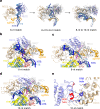
Fig. 1. Target DNA binding induces Cas9 REC lobe restructuring.
a, Top, schematic depicting DNA-bound complexes with increasing extent of complementarity to guide RNA. Bottom, domain composition of SpCas9. 1-A, REC1-A domain; I–III, RuvC domain motifs I–III; BH, bridge helix. b, Structural comparison of the SpCas9 binary (left), 6-nt match (middle) and 8-nt match (right) complexes. c, Zoomed-in view of the seed region of the guide RNA–target DNA heteroduplex in the 6-nt match complex. Tyr450 stacks between the fifth and sixth nucleotide, counting from the PAM-proximal end of the heteroduplex. d, Zoomed-in view of the seed region of the guide RNA–target DNA heteroduplex in the 8-nt match complex. e, Fitted cleavage rate (kobs) of wild-type (WT) and Y450A mutant Cas9 against on-target and off-target substrates. Data represent mean fit ± s.e.m. of n = 4 independent replicates. Two-tailed t-test, ****P < 0.0001, ***P = 0.0002, **P = 0.0011. The P-value for the on-target dataset was not significant (P = 0.1058). Source data
Fig. 2
Fig. 2. R-loop propagation drives DNA repositioning within Cas9.
a, Zoomed-in views of the conformational transitions in the PAM-distal DNA duplex and Cas9 REC2 and REC3 domains in the 6-, 8- and 10-nt match complex. b, Zoomed-in view of the R-loop in the 6-nt match complex. c, Zoomed-in view of the R-loop in the 8-nt match complex. d, Zoomed-in view of the R-loop in the 10-nt match complex. e, Zoomed-in view of the interaction between the REC2 domain DDD helix and the REC3 RRR helix.
Fig. 3
Fig. 3. Target pairing past the seed region undocks the HNH nuclease domain.
a, Position of the HNH catalytic site in the binary, 6-, 8- and 10-nt match complexes. b, Structural overlay of the REC2 and REC3 domains in the 10-, 12-, 14-, 16- and 18-nt match (checkpoint) complexes. c, Overview of the 12-nt match (left), 14-nt match (middle) and 16-nt match (right) complexes, shown in the same orientations. For each complex, the unsharpened cryo-EM map is overlaid with the respective atomic model. The 12-nt match complex map shows residual density for the displaced NTS (white). The 14-nt match map reveals residual density corresponding to the HNH domain. No density is visible for NTS. Cryo-EM maps are coloured according the schematic in Fig. 1a.
Fig. 4
Fig. 4. HNH domain rotation and DNA bending enable catalytic activation.
a, The structure of the 18-nt match complex in the pre-cleavage, checkpoint state. b, The structure of the 18-nt match complex in the catalytically active state. c, Conformations of the guide–target heteroduplexes and REC2 and REC3 domains in the 18-nt match checkpoint (left) and catalytic (right) complexes. The structures are shown in the same orientations as in a,b. The HNH domain has been omitted from the images for clarity. d, Zoomed-in view of the HNH nuclease active site in the 18-match catalytic complex containing bound cleaved TS. e, Zoomed-in view of the L1 linker contacting the minor groove of the guide RNA–target DNA heteroduplex. f, Zoomed-in view of the RuvC nuclease active site containing the 3′-terminal product of cleaved NTS.
Extended Data Fig. 1
Extended Data Fig. 1. Minimal target complementarity necessary for stable Cas9 binding.
Size exclusion chromatography analysis of nuclease-inactive SpCas9 complexed with sgRNA and Cy5-labeled DNA substrates with increasing extent of guide-target complementarity. A254/A280 and Cy5 signals were normalised.
Extended Data Fig. 2
Extended Data Fig. 2. Schematic representation of DNA substrates used in structural studies.
Base pair complementarity between sgRNA, target strand (TS), and non-target strand (NTS) is indicated by black lines.
Extended Data Fig. 3
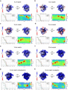
Extended Data Fig. 3. Cryo-EM density maps of DNA-bound SpCas9 complexes.
Front (left) and back (right) views of unsharpened cryo-EM density maps of the partially-bound SpCas9 complexes. Maps are coloured by local resolution, and gold-standard FSC of 0.143 resolution graphs and particle distribution heatmaps are indicated for each complex.
Extended Data Fig. 4
Extended Data Fig. 4. Structural models of DNA-bound SpCas9 complexes.
Cartoon representations of DNA-bound 6-, 8-, 10-, 12-, 14-, 16-, 18-nt match (checkpoint and catalytic) complexes of SpCas9. Each model was generated based on the corresponding map shown in Extended Data Fig. 3.
Extended Data Fig. 5
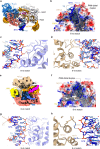
Extended Data Fig. 5. Stabilisation of the PAM-distal duplex by REC2/3 domains.
a, Structural overlays of the SpCas9 bridge helix (BH), REC1, RuvC, and PAM-interacting (PI) domains, as well as the PAM-proximal DNA duplex and the sgRNA from the partially bound complex structures determined by crystallography and cryoEM, and full R-loop complexes (PDB: 6O0X, 6O0Y, 6O0Z). b, Zoom-in view of the PAM-distal DNA duplex in the 6-nt match complex. The protein surface is coloured according to electrostatic surface potential, with red denoting negative and blue positive charge. c, Interactions between SpCas9 REC2 domain and the backbone of the PAM-distal NTS in the 6-nt match complex. d, Interactions between the REC3 domain and the backbone of the PAM-distal TS in the 6-nt match complex. e, Central slice through the 6-nt match complex. Cryo-EM density map is coloured according to Fig. 1a. White density indicates positioning of the 5’ sgRNA end. f, PAM-distal DNA duplex in the 8-nt match complex remains positioned in a positively charged cavity between the REC2 and REC3 domains. The protein surface is coloured according to electrostatic surface potential. g, Interactions of the REC2 domain with the PAM-distal DNA duplex in the 8-nt match complex. h, Interactions of the REC3 domain with the NTS of the PAM-distal duplex in the 8-nt match complex.
Extended Data Fig. 6
Extended Data Fig. 6. In vitro cleavage activities of structure-guided REC2 and REC3 mutants of Cas9.
a, Off-target sequences selected for nuclease activity assays. Nucleotide mismatches between the TRAC guide RNA and the target are highlighted; matching nucleotides are denoted by a dot. b, In vitro cleavage kinetics of Y450A mutants from which kobs values are derived using single exponential fitting. Data represents mean ± SEM (n = 4). c, In vitro cleavage kinetics of REC2/REC3 mutants from which kobs values are derived using single exponential fitting. Data represents mean ± SEM (n = 4). d, Cleavage rate constants of PAM-distal duplex stabilising REC2/REC3 mutants on on- and off-targets. Data represents mean fit ± SEM of n = 4 replicates, significance was determined by a two-tailed t-test. *, p < 0.05; **, p < 0.01; ***, p < 0.001; ****, p < 0.0001. e, In vitro cleavage kinetics of DDD and RRR helix mutants from which kobs values are derived using single exponential fitting. Data represents mean ± SEM (n = 4). f, Cleavage rate constants of Cas9 DDD and RRR helix mutants. Data represents mean fit ± SEM of n = 4 replicates, significance was determined by a two-tailed t-test. *, p < 0.05; **, p < 0.01; ***, p < 0.001; ****, p < 0.0001. On- and off-target substrates were fluorescently labelled on the PAM-proximal end of the target DNA strand in all panels. Source data
Extended Data Fig. 7
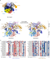
Extended Data Fig. 7. PAM-distal positioning and REC lobe conformation in the 10-nt match complex.
a, Cryo-EM density of the 10-nt match complex overlaid with the structural model. NTS density can be traced along the heteroduplex (white). b, Cartoon representations of the X-ray crystallographic structures of the 10-nt match complex as based on the two complex copies (molecules A and B) in the crystallographic asymmetric unit. The complexes exhibit highly similar conformations (RMSD 0.46 Å). c, Alignment of protein sequences of the REC2 DDD and REC3 RRR helices from REC2-containing Cas9 orthologs.
Extended Data Fig. 8
Extended Data Fig. 8. HNH undocking induced by R-loop extension.
a, Residual HNH domain density (white) observed in the 14-nt match complex, in which the elongated heteroduplex establishes a contact with the L2 linker. No NTS density is observed past the PAM region due to disorder. b, Zoom-in view of the interaction between the HNH domain L2 linker and the RuvC 1030–1040 helix induced by heteroduplex proximity of the 16-bp complex. c, HNH domain relocation towards the binding channel results in the formation of a positively charged NTS binding channel. No residual electron density (white) is observed for the NTS in the absence of the PAM-distal duplex. The protein is coloured according to electrostatic surface potential, with red being negative, blue positive. d, Surface electrostatics map of the 18-nt match catalytic state of SpCas9, showing the NTS binding cleft with cleaved NTS positioned within the active site.
Extended Data Fig. 9
Extended Data Fig. 9. Molecular mechanism of Cas9 R-loop formation and conformational activation.
In the RNA-bound (binary) complex, the central DNA binding channel is occluded by the REC2 and REC3 domains. Upon PAM recognition and initial 5-nt base pairing with the seed sequence of the guide RNA, the REC2 domain is displaced to form a binding cleft to accommodate the PAM-distal DNA duplex. Formation of 8-bp heteroduplex further displaces the REC3 domain and fully opens the central binding channel, while the PAM-distal duplex remains in the REC2/3 cavity. Extension of the R-loop to 10-bp heteroduplex places the guide-TS heteroduplex and the PAM-distal duplex into the central binding channel, accompanied by formation of electrostatic contacts between the REC2 and REC3 domains. Base pairing past the seed region results in undocking of the HNH domain from the RuvC and PI domain interface, and results in its repositioning towards target heteroduplex into the checkpoint state. R-loop formation past 17 base pairs induces REC2 domain displacement from the binding channel and rotation of the HNH domain active site towards the TS cleavage site, while simultaneously positioning the NTS in the RuvC domain active site.

Similar articles

Cited by

References

    1. Garneau JE, et al. The CRISPR/Cas bacterial immune system cleaves bacteriophage and plasmid DNA. Nature. 2010;468:67–71. doi: 10.1038/nature09523. - DOI - PubMed
    1. Jinek M, et al. A programmable dual-RNA-guided DNA endonuclease in adaptive bacterial immunity. Science. 2012;337:816–821. doi: 10.1126/science.1225829. - DOI - PMC - PubMed
    1. Sapranauskas R, et al. The Streptococcus thermophilus CRISPR/Cas system provides immunity in Escherichia coli. Nucleic Acids Res. 2011;39:9275–9282. doi: 10.1093/nar/gkr606. - DOI - PMC - PubMed
    1. Cong L, et al. Multiplex genome engineering using CRISPR/Cas systems. Science. 2013;339:819–823. doi: 10.1126/science.1231143. - DOI - PMC - PubMed
    1. Jinek M, et al. RNA-programmed genome editing in human cells. eLife. 2013;2:e00471. doi: 10.7554/eLife.00471. - DOI - PMC - PubMed