. 2023 Apr;40(4):801-816.
doi: 10.1007/s11095-022-03350-7. Epub 2022 Aug 24.

Biophysical, Molecular and Proteomic Profiling of Human Retinal Organoid-Derived Exosomes

Affiliations

Biophysical, Molecular and Proteomic Profiling of Human Retinal Organoid-Derived Exosomes

Peggy Arthur et al. Pharm Res. 2023 Apr.

Abstract

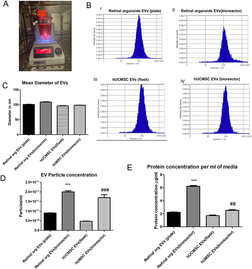
Purpose: There is a growing interest in extracellular vesicles (EVs) for ocular applications as therapeutics, biomarkers, and drug delivery vehicles. EVs secreted from mesenchymal stem cells (MSCs) have shown to provide therapeutic benefits in ocular conditions. However, very little is known about the properties of bioreactor cultured-3D human retinal organoids secreted EVs. This study provides a comprehensive morphological, nanomechanical, molecular, and proteomic characterization of retinal organoid EVs and compares it with human umbilical cord (hUC) MSCs.

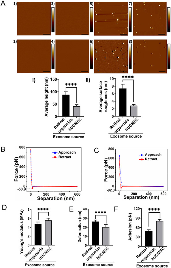
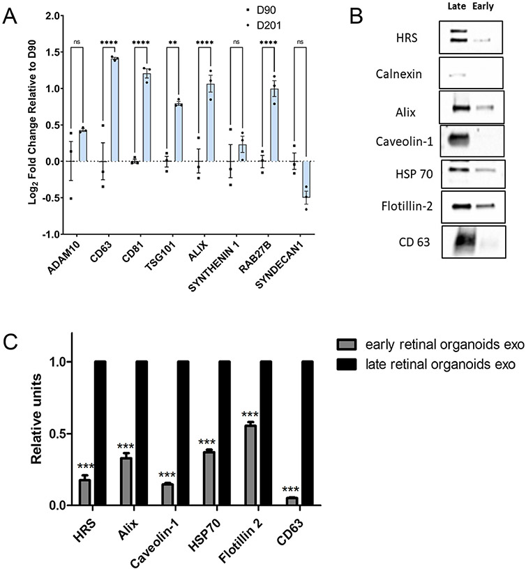
Methods: The morphology and nanomechanical properties of retinal organoid EVs were assessed using Nanoparticle tracking analysis (NTA) and Atomic force microscopy (AFM). Gene expression analysis of exosome biogenesis of early and late retinal organoids were compared using qPCR. The protein profile of the EVs were analyzed with proteomic tools.

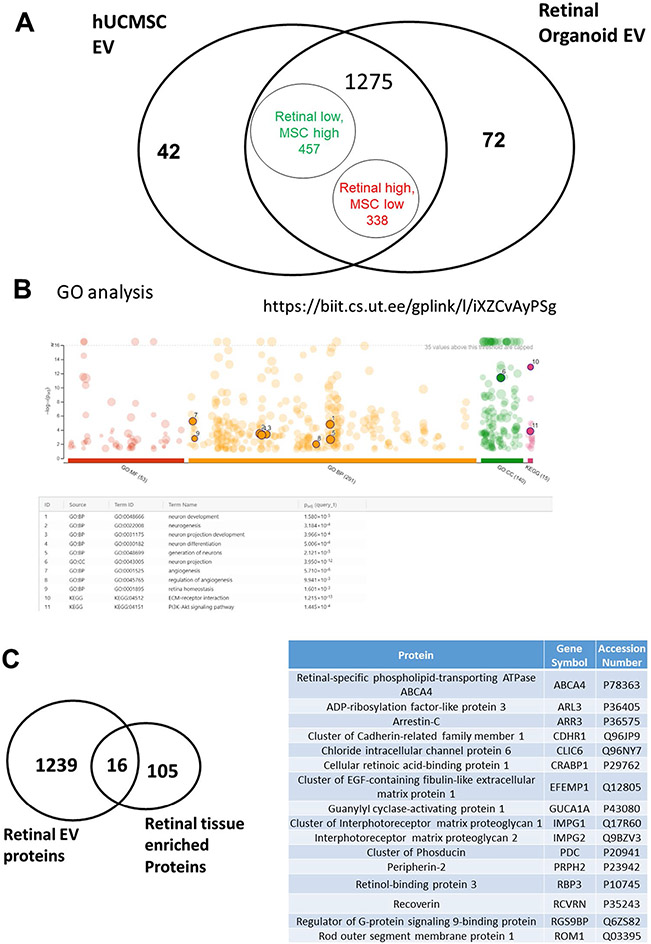
Results: NTA indicated the average size of EV as 100-250 nm. A high expression of exosome biogenesis genes was observed in late retinal organoids EVs. Immunoblot analysis showed highly expressed exosomal markers in late retinal organoids EVs compared to early retinal organoids EVs. Protein profiling of retinal organoid EVs displayed a higher differential expression of retinal function-related proteins and EV biogenesis proteins than hUCMSC EVs, implicating that the use of retinal organoid EVs may have a superior therapeutic effect on retinal disorders.

Conclusion: This study provides supplementary knowledge on the properties of retinal organoid EVs and suggests their potential use in the diagnostic and therapeutic treatments for ocular diseases.

Keywords: Bioreactor; Extracellular vesicles; Human retinal organoids; Proteomics; Stem cells.


Figures

Fig. 1
Fig. 1
Retinal organoids differentiation and characterization. (A) Schematic illustration showing the differentiation of retinal organoids from hiPSC and timeline representation of the early (Day 30–120) and late organoids (Day 121–300) for exosome preparation. (B) Phase contrast microscopic images showing the morphology of retinal organoids at different time points of differentiation (Day 2, B1; Day 25, B2; Day 45, B3; Day 200, B4). Scale bar, 200 μm. (C) Confocal images of 90-day old retinal organoids (C1-8) stained for retinal progenitor cells (Pax6, C1; Vx2, C2), ganglion and amacrine cells (Brn3a, C3), pan photoreceptors (Crx, C4; Rcvrn, C5; Aipl1; C6), rod photoreceptor (NRL; C7), and cone photoreceptor (ARR3, C8). Image of 200-day old retinal organoids showing the staining of rhodopsin protein indicating the maturation of rod photoreceptor (Rho, C9). Scale bar, 50 μm.
Fig. 2
Fig. 2
Characterization of Retinal organoid derived EVs by NTA. A Retinal organoids in PBS VW bioreactor. B Comparison of EVs from retinal organoids (plate and bioreactor) and hUCMSC (flask and bioreactor) culture. Representative NTA histogram for i) retinal organoids EVs (plate) ii) retinal organoids EVs (bioreactor) iii) hUCMSC EVs (flask) iv) hUCMSC EVs (bioreactor). C Mean diameter of EVs in nm. D Retinal organoid and hUCMSCs EV particle concentration per mL of PBS E Protein content per mL of conditioned medium (* retinal organoid EVs(plate vs bioreactor), # hUCMSC EVs(flask vs bioreactor), *,# indicate p < 0.05, **, ## indicate p < 0.01 and ***, ### p < 0.001.
Fig. 3
Fig. 3
Topographical and nanomechanical characteristics of EVs. A Representative AFM images of plain mica and APTES: DIPEA modified mica (1, 3) Height image (2) Peak force error image. Representative images of retinal organoid and hUCMSC EVs (5, 7) Height image 6, 8) Peak force error image. (Scale bar: 1 μm; color bar for height image: −65 nm to 65 nm; color bar for peak force error image: −300 pN to 300 pN). Morphology quantification i) Average height ii) Average surface roughness. (Statistical significance: ****; p < 0.0001). A representative force-separation curve displaying adhesion value corresponding to exosomes derived from B Retinal organoid and C hUCMSC. Nanomechanical attributes displaying D Young’s modulus E Deformation F Adhesion. (****; p < 0.0001).
Fig. 4
Fig. 4
Biogenesis of exosome in human 3D retinal organoids. A qRT-PCR assay of early (Day 90) and late (Day 200) retinal organoids showing the expression of ESCRT-dependent (Alix, TSG101), ESCRT-independent (Synthenin1, Syndecan1), Rabs family of transport and membrane fusion and pan exosome targets (Rab27B, CD63, CD81, ADAM10). (ns, p > 0.05; *p ≤ 0.05; **p ≤ 0.01; ***p ≤ 0.001; ****p ≤ 0.0001). B Western blots of early and late retinal organoid EVs for exosome markers HRS, Calnexin, Alix, Caveolin 1, HSP 70, Flotillin-2 and CD63. C Densitometric analysis of western blots. ***represents p < 0.001.
Fig. 5
Fig. 5
Proteomics analysis comparing retinal organoid with hUCMSC EVs. A Venn diagram of total identified proteins in both EV groups. B GO analysis of Retinal EVs. C Venn diagram of retinal organoid EVs, Human Protein ATLAS of retinal tissue and overlapped proteins between them.

Similar articles

Cited by

References

    1. Hartong DT, Berson EL, Dryja TP. Retinitis pigmentosa. The Lancet. 2006;368(9549):1795–809. - PubMed
    1. Goodwin P. Hereditary retinal disease. Curr Opin Ophthalmol. 2008;19(3):255–62. - PubMed
    1. Hamel J-F. Les ruines du progrès chez Walter Benjamin: anticipation futuriste, fausse reconnaissance et politique du présent. Protée. 2007;35(2):7–14.
    1. Kutlehria S, Sachdeva MS. Role of in vitro models for development of ophthalmic delivery systems. Crit Rev Ther Drug Carrier Syst. 2021;38(3). - PMC - PubMed
    1. Buskin A, Zhu L, Chichagova V, Basu B, Mozaffari-Jovin S, Dolan D, Droop A, Collin J, Bronstein R, Mehrotra S. Disrupted alternative splicing for genes implicated in splicing and ciliogenesis causes PRPF31 retinitis pigmentosa. Nat Commun. 2018;9(1):1–19. - PMC - PubMed