Review
. 2022 Jun;15(6):1143-1157.
doi: 10.1038/s41385-022-00551-6. Epub 2022 Aug 24.

Mucosal immunology of the ocular surface

Affiliations
Review

Mucosal immunology of the ocular surface

Cintia S de Paiva et al. Mucosal Immunol. 2022 Jun.

Abstract

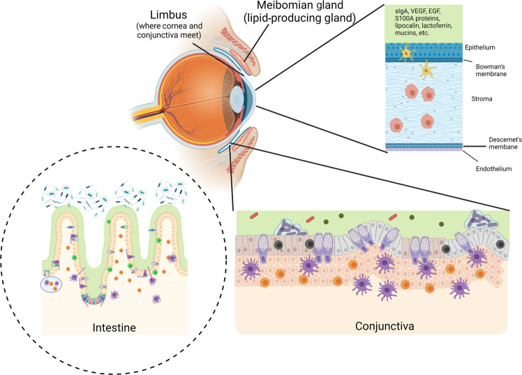
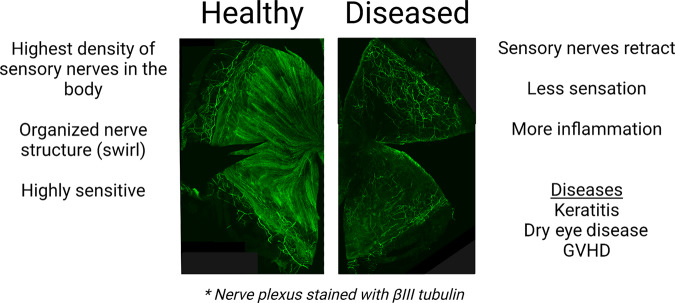
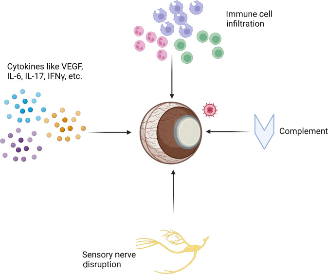
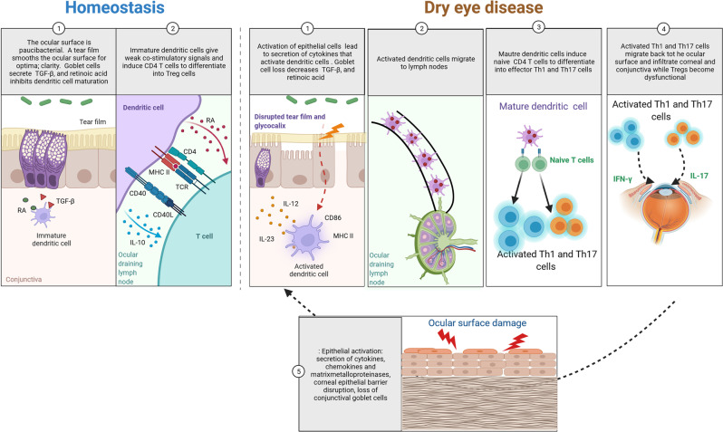
The eye is a sensory organ exposed to the environment and protected by a mucosal tissue barrier. While it shares a number of features with other mucosal tissues, the ocular mucosal system, composed of the conjunctiva, Meibomian glands, and lacrimal glands, is specialized to address the unique needs of (a) lubrication and (b) host defense of the ocular surface. Not surprisingly, most challenges, physical and immunological, to the homeostasis of the eye fall into those two categories. Dry eye, a dysfunction of the lacrimal glands and/or Meibomian glands, which can both cause, or arise from, sensory defects, including those caused by corneal herpes virus infection, serve as examples of these perturbations and will be discussed ahead. To preserve vision, dense neuronal and immune networks sense various stimuli and orchestrate responses, which must be tightly controlled to provide protection, while simultaneously minimizing collateral damage. All this happens against the backdrop of, and can be modified by, the microorganisms that colonize the ocular mucosa long term, or that are simply transient passengers introduced from the environment. This review will attempt to synthesize the existing knowledge and develop trends in the study of the unique mucosal and immune elements of the ocular surface.


Conflict of interest statement

The authors declare no competing interests.

Figures

Fig. 1
Fig. 1. Similarities and differences between intestinal and ocular surface mucosal immune tissues.
Similarities between the intestinal and ocular surface mucosal immune tissues include being continually bathed in mucus, harboring a diverse array of immune cells, the presence of goblet cells, and associations with bacteria. Differences include the presence of Peyer’s patches (light blue oval) in the intestines, which are not found in the conjunctiva. Also, the types, amount, and diversity of bacteria associated with the tissues is vastly different. Goblet cells (purple cells in the epithelial layer of the conjunctiva and intestines) are more concentrated in the conjunctiva as compared to the intestines. The meibomian glands associated with the eyelid supply the lipid fraction of the tears that coat the ocular surface. While both tissues are bathed in mucus, MUC5A is the tear associated mucin while MUC2 is associated with intestinal mucus. Tears contain a diverse array of factors that prevent microbes from adhering and infecting the ocular surface including: secretory IgA (sIgA), VEGF, EGF, S100A proteins, lipocalin, lactoferrin, and other miscellaneous factors.
Fig. 2
Fig. 2. Ocular surface diseases can lead to a loss of sensory nerves in the cornea.
In a normal corneal, thick sensory nerve trunk begin at the limbus (peripheral to the cornea) and innervate the cornea eventually thinning to become fine sensory nerve endings which terminate in the corneal epithelium. Fine sensory nerve endings become more concentrated and begin to swirl as they reach the central cornea (left). During ocular surface disease, sensory nerve endings can retract (right) and this results in a loss of blink reflex that can be temporary or permanent. Loss of blink reflex can leave the eye susceptible to infection, dry eye phenotypes and excessive inflammation. Nerves in the figure were stained with βIII tubulin.
Fig. 3
Fig. 3. Factors that contribute to Herpes Stromal Keratitis (HSK).
After a predominantly sub-clinical primary infection, HSV-1 gains access to sensory neurons and enters a latent state within trigeminal ganglion neurons indefinitely. Spontaneously or during times of immune suppression, HSV-1 can reactivate and virions can be deposited at the cornea resulting in an inflammatory response. Repeated reactivation events result in: (1) increased recruitment of inflammatory immune cells, (2) the production of inflammatory cytokines/chemokines, (3) disruption of sensory nerves, and (4) the initiation of complement, which preferentially targets sensory nerves. Because HSK is a multi-factoral disease, these processes all contribute to disease and ultimately result in tissue damage, fibrosis, and eventual blindness.
Fig. 4
Fig. 4. The immune response in dry eye disease and the vicious cycle of inflammation.
Homeostasis: 1-In homeostasis, the ocular surface is paucibacterial and has innumerous anti-microbial defenses, including a healthy tear film rich in anti-microbial proteins (SA100s, lysozyme). In the conjunctiva, goblet cells secrete retinoic acid (RA) and TGF-beta that maintain the resident dendritic cells in an immature state. 2- Immature dendritic cells provide ocular surveillance and migrate constantly to the ocular draining nodes where regulatory T cells (Tregs) are preferentially primed. dry eye disease: 1-Insults to the ocular surface cause activation of epithelial cells, disrupted tear film and glycocalyx which in turn lead to secretion of chemokines, secretion of T helper (Th) polarizing cytokines such as IL-12 and IL-23 and upregulation of CD86 and MHC II, leading to activated dendritic cells. Loss of goblet cells and squamous metaplasia of the conjunctival epithelium lead to goblet cells that do not properly secrete at the ocular surface. Secondary to loss of goblet cells, there is less RA and TGF-β, leading to further APC activation. 2-Activated dendritic cells migrate to ocular lymph nodes. 3-Mature dendritic cells primer naïve T cells into Th1 and Th17 cells. 4-Activated Th1 and Th17 cells migrate back to the eye, where they secrete IFN-γ and IL-17, which cause goblet cell loss, corneal barrier disruption and activation of innate immunity. Dysfunctional Tregs also participate in the immune response of dry eye disease (not despicted). 5- Insults to the ocular surface reinitiate the vicious cycle of dry eye activating epithelial cells, disrupting the tear film and glycocalyx which in turn lead to secretion of chemokines, secretion of Th polarizing cytokines such as IL-12 and IL-23 and upregulation of CD86 and MHC II, leading to activated dendritic cells. Loss of goblet cells and squamous metaplasia of the conjunctival epithelium lead to goblet cells that do not properly secrete at the ocular surface. Secondary to loss of goblet cells, there is less RA and TGF-β, leading to further APC activation.

References

    1. Sridhar MS. Anatomy of cornea and ocular surface. Indian J. Ophthalmol. 2018;66:190–194. doi: 10.4103/ijo.IJO_646_17. - DOI - PMC - PubMed
    1. Xiao Y, et al. Goblet cell-produced retinoic acid suppresses CD86 expression and IL-12 production in bone marrow-derived cells. Int. Immunol. 2018;30:457–470. doi: 10.1093/intimm/dxy045. - DOI - PMC - PubMed
    1. Contreras-Ruiz L, Ghosh-Mitra A, Shatos MA, Dartt DA, Masli S. Modulation of conjunctival goblet cell function by inflammatory cytokines. Mediators Inflamm. 2013;2013:636812. doi: 10.1155/2013/636812. - DOI - PMC - PubMed
    1. Payne AP. The harderian gland: a tercentennial review. J. Anat. 1994;185:1–49. - PMC - PubMed
    1. Knop E, Knop N. Lacrimal drainage-associated lymphoid tissue (LDALT): a part of the human mucosal immune system. Invest Ophthalmol. Vis. Sci. 2001;42:566–574. - PubMed

Publication types