Review
. 2022 Aug 24;17(1):205.
doi: 10.1186/s13019-022-01962-z.

Rare ectopic thyroid tissue as multiple bilateral pulmonary nodules: a case report and literature review

Affiliations
Review

Rare ectopic thyroid tissue as multiple bilateral pulmonary nodules: a case report and literature review

Jianfeng Tan et al. J Cardiothorac Surg. .

Abstract

Background: The prevalence of ectopic thyroid tissue is 1 in every 100,000 to 300,000 persons in the general population, and ectopic thyroid tissue in the bilateral lung lobes is even rarer. Due to its rarity, there is no definitive or standard guidance on the diagnosis and treatment of ectopic thyroid tissue presenting as multiple bilateral pulmonary nodules.

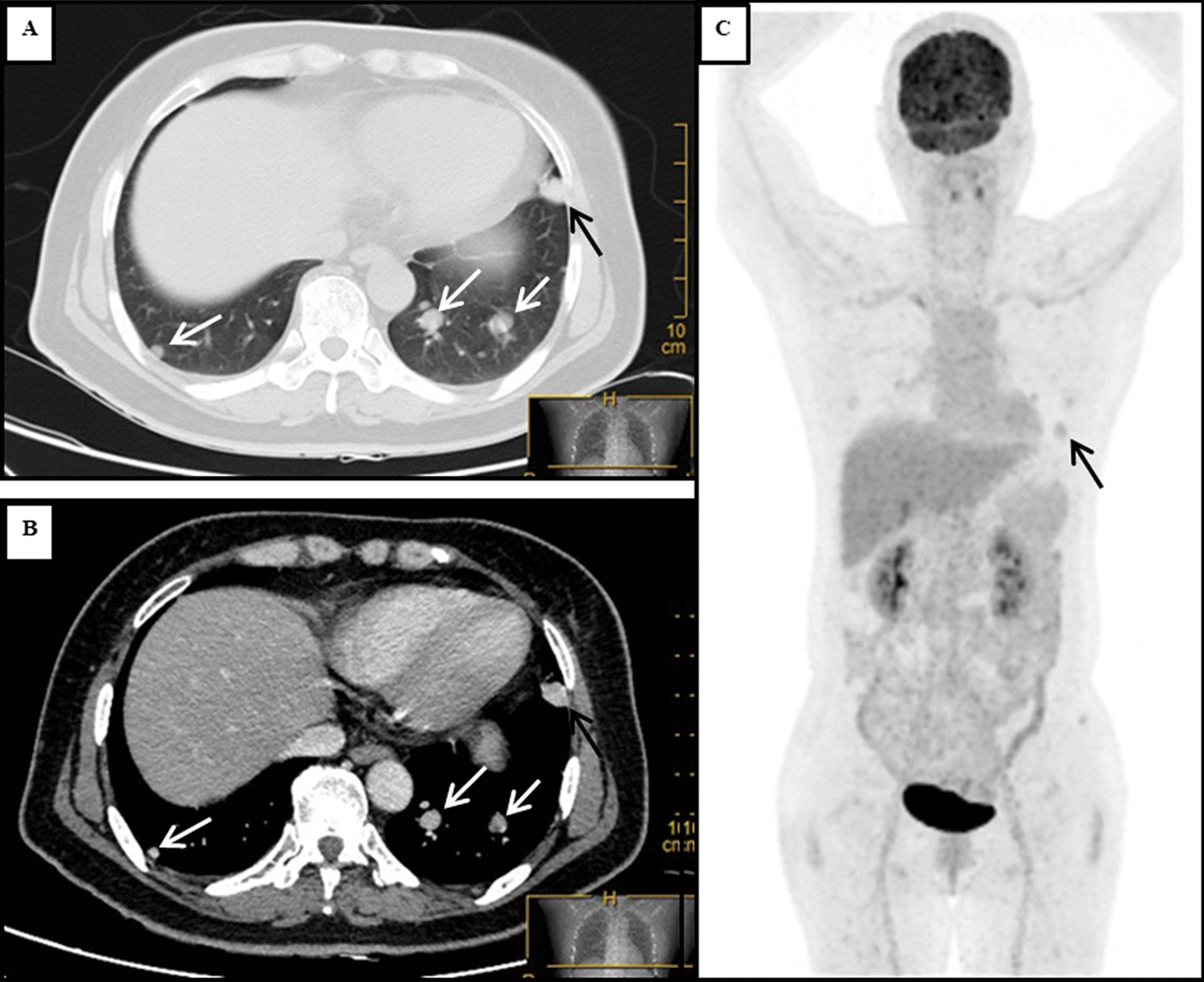
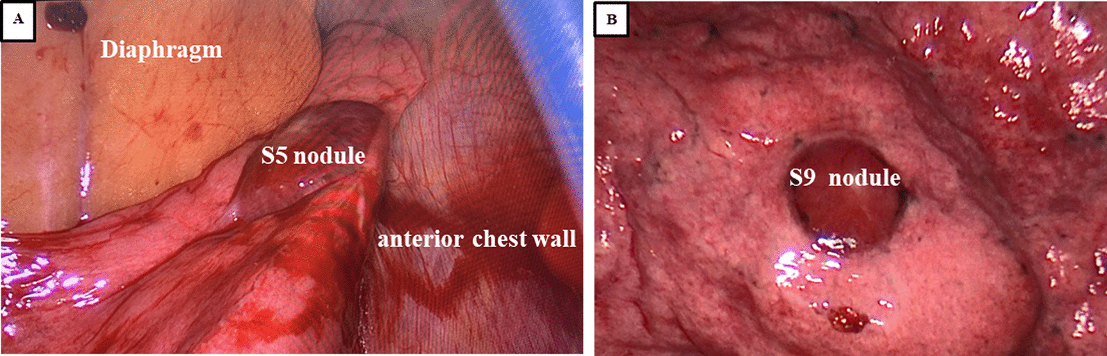
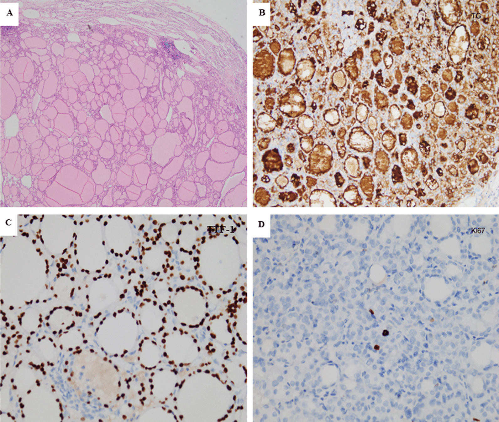
Case presentation: A 56-year-old woman presented with multiple bilateral pulmonary nodules, and the patient had a history of hyperthyroidism but had no symptoms of ectopic thyroid tissue. Computed tomography (CT) demonstrated multiple solid nodules in both lungs, and the largest nodule (sized 15 × 14 mm) was located in segment 5 of the upper left lung. The initial diagnosis based on imaging was metastatic malignancies. Positron emission tomography-computed tomography (PET-CT) showed multiple bilateral intrapulmonary nodules that had slightly increased metabolism (SUVmax 1.7). The largest pulmonary nodule and another nodule in the left lung were resected by video-assisted thoracoscopy surgery (VATS). The pathological and immunohistochemical (IHC) examinations confirmed a diagnosis of ectopic thyroid tissue. No postoperative adjuvant therapy was given, and the patient was discharged 3 days after the operation and had regular follow-up examinations.

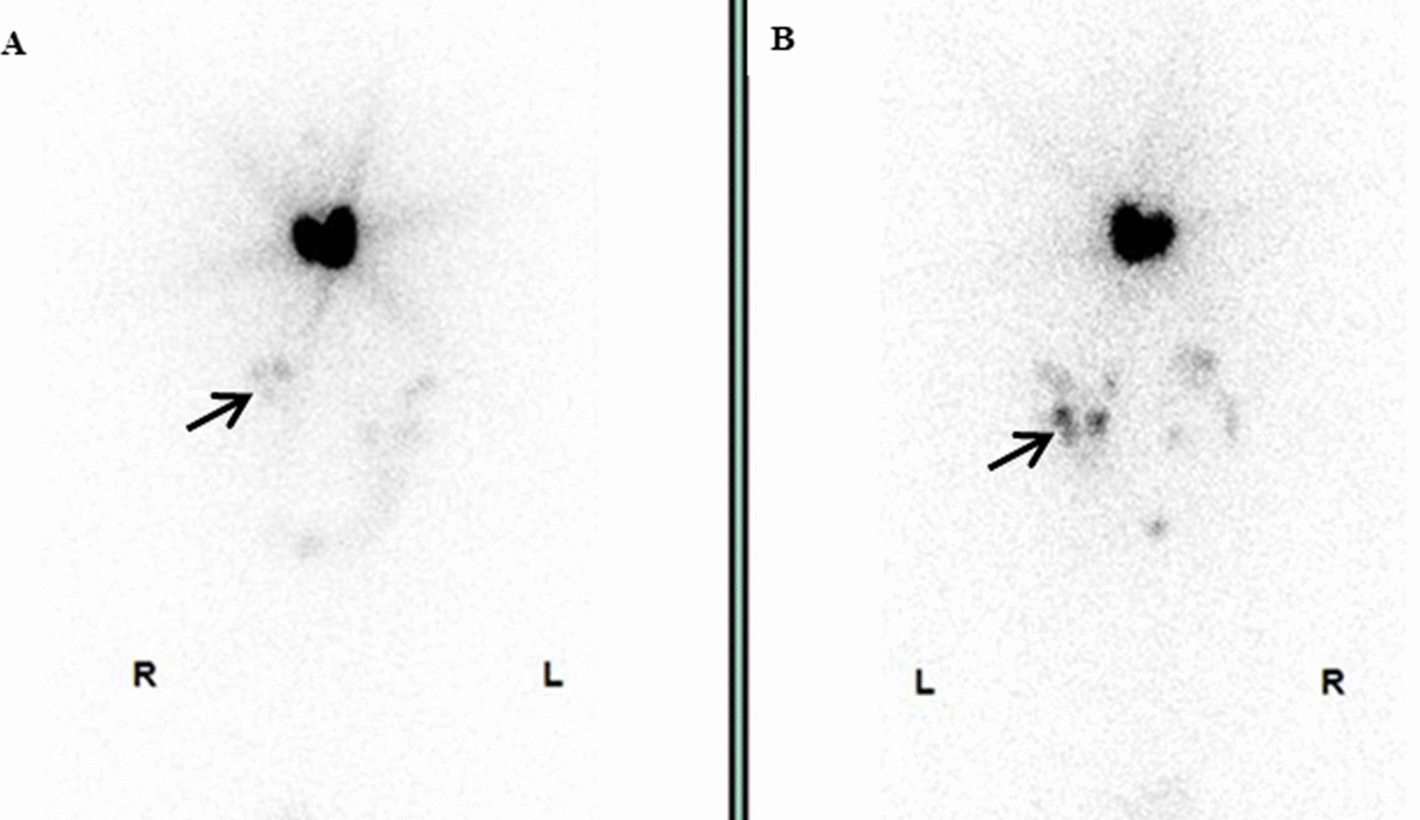
Conclusion: The diagnosis of ectopic thyroid tissue in the bilateral lung lobes is extremely difficult and should be considered carefully. PET-CT and surgical resection of intrapulmonary nodules are alternatives for clinicians in diagnosing ectopic thyroid tissue. Regular postoperative follow-up is needed.

Keywords: Case report; Ectopic thyroid; Pulmonary nodules; VATS.


Conflict of interest statement

The authors declare that they have no competing interests.

Figures

Fig. 1
Fig. 1
The chest CT scan and PET-CT examination showed multiple bilateral nodules in the patient’s lung lobes. The lung window (A) and longitudinal window (B) demonstrated that some round, well-defined and various-sized nodules were seen in both lungs (white arrow), the largest nodule was found in segment 5, and that nodule measured 15*14 mm (black arrow). (C) PET-CT indicated that there were multiple nodules in the bilateral lung lobes with mildly increased metabolism and that there was a larger nodule in the left lung (black arrow)
Fig. 2
Fig. 2
Intraoperative exploration revealed multiple nodules in the left lung that were dark red and soft, and these were protruding from the visceral pleura. The segment 5 (S5) nodule (A) and segment 9 (S9) nodule (B) were resected
Fig. 3
Fig. 3
Microphotograph of the patient’s thyroid ectopia in the left lung lobes. A Haematoxylin and eosin (H&E) staining showed that the pulmonary nodules consisted of typical thyroid tissue with follicles containing colloids (magnification × 40). B Immunohistochemistry staining of the follicular epithelial cells showed positivity for thyroglobulin (TG, magnification ×100). C Positive immunohistochemical staining for thyroid transcription factor-1 (TTF-1, magnification ×100). D Immunohistochemistry staining of follicular epithelial cells showed weak positivity for Ki67 (< 1%, magnification × 100)
Fig. 4
Fig. 4
I-131 scintigraphy, anterior A and posterior B views showed that functional thyroid tissue had developed in the patient’s neck and that the multiple nodular radioactive distributions that were in the bilateral lung lobes were concentrated (black arrow)

Similar articles

Cited by

References

    1. Noussios G, Anagnostis P, Goulis DG, Lappas D, Natsis K. Ectopic thyroid tissue: anatomical, clinical, and surgical implications of a rare entity. Eur J Endocrinol. 2011;165:375–382. doi: 10.1530/EJE-11-0461. - DOI - PubMed
    1. Weng LP, Kaiser A, Koch HH. Wünsch PH [Multilocular thyroid gland ectopy] Dtsch Med Wochenschr. 2000;125:421–424. doi: 10.1055/s-2007-1024233. - DOI - PubMed
    1. Gu YM, Fan YY, Wang WP, Chen LQ. Ectopic thyroid tissue in the heart. QJM. 2019;112:927–928. doi: 10.1093/qjmed/hcz219. - DOI - PubMed
    1. Chen M, Hu J, Cai X. Ectopic thyroid gland tissue in the liver. Clin Gastroenterol Hepatol. 2020;18:e157. doi: 10.1016/j.cgh.2019.09.042. - DOI - PubMed
    1. Li Y, Li S, Wang M, Tong L. Ectopic thyroid in the gallbladder accompanied with gallbladder adenoma: a case report. Medicine (Baltimore) 2019;98:e18293. doi: 10.1097/MD.0000000000018293. - DOI - PMC - PubMed

MeSH terms