. 2022 Aug 24;19(1):208.
doi: 10.1186/s12974-022-02569-w.

Psychological stress induces depressive-like behavior associated with bone marrow-derived monocyte infiltration into the hippocampus independent of blood-brain barrier disruption

Affiliations

Psychological stress induces depressive-like behavior associated with bone marrow-derived monocyte infiltration into the hippocampus independent of blood-brain barrier disruption

Huiling Hu et al. J Neuroinflammation. .

Abstract

Background: Psychological stress is one of the most important factors that trigger emotional disorders, such as depression and anxiety. Emerging evidence suggests that neuroinflammation exacerbated by bidirectional communication between the peripheral immune system and the central nervous system facilitates abnormal psychiatric symptoms. This study aimed to investigate the hippocampal migration of bone marrow (BM)-derived monocytes and its role in regulating depressive-like behaviors using the chronic psychological stress (CPS) mouse model. More importantly, whether the central migration of these peripheral BM-derived cells depend on the disruption of the blood-brain barrier (BBB) was also investigated.

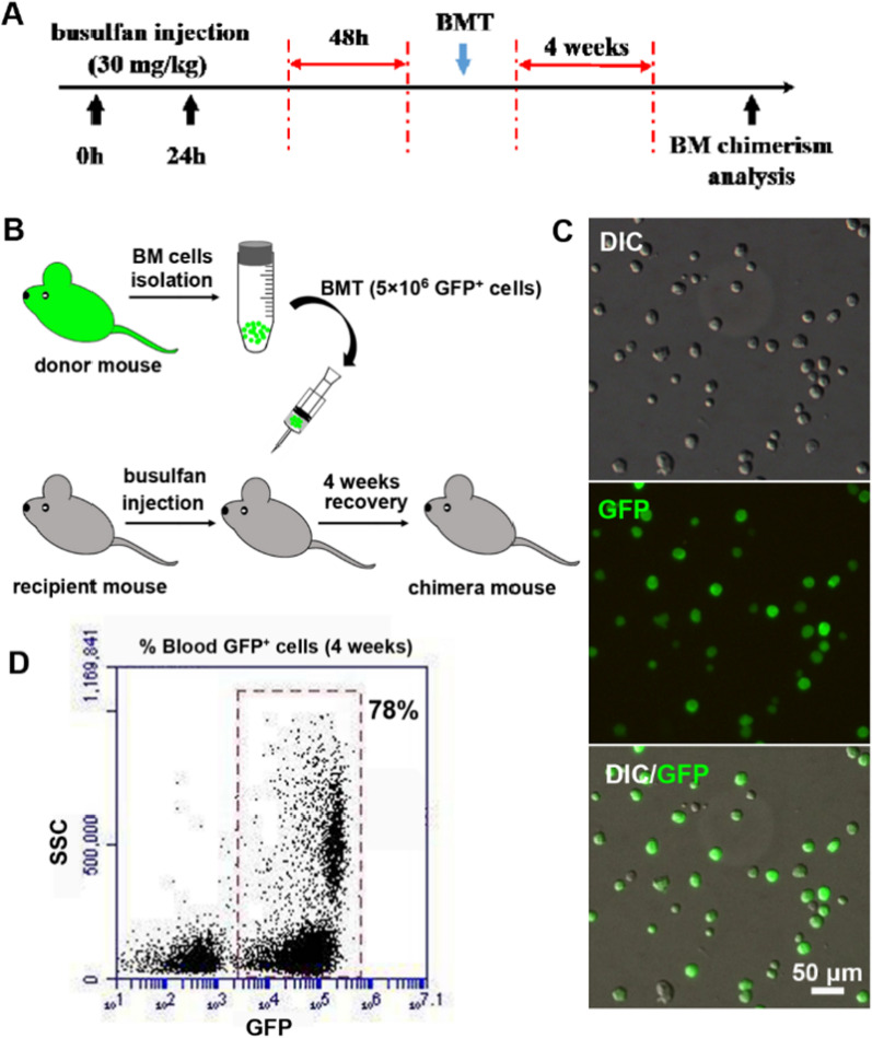
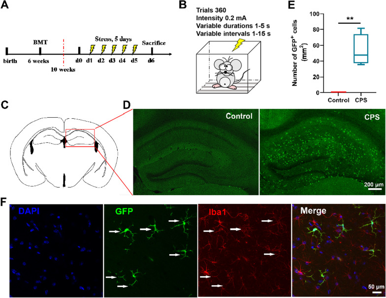
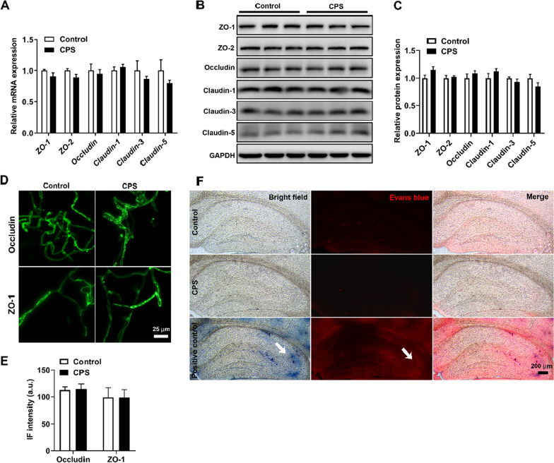
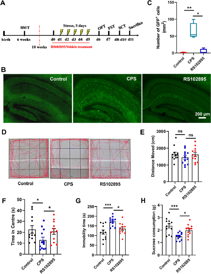
Methods and findings: Green fluorescent protein-positive (GFP+) BM chimeric mice were used to distinguish BM-derived monocytes within the brain. A CPS mouse model was established to explore the effect of CPS on hippocampal migration of BM-derived monocytes and its role in the regulation of depressive-like behaviors. The results revealed that BM-derived GFP+ cells accumulated in the hippocampus and differentiated into microglia-like cells after exposure to CPS. Interestingly, this migration was not associated with BBB disruption. Furthermore, treatment with C-C chemokine receptor 2 (CCR2) antagonist (RS102895) suppressed the recruitment of BM-derived monocytes to the hippocampus and alleviated depressive-like symptoms.

Conclusion: These findings indicate that monocyte recruitment to the hippocampus in response to psychological stress may represent a novel cellular mechanism that contributes to the development of depression.

Keywords: Blood–brain barrier; Bone marrow transplantation; Depression; Hippocampus; Monocytes; Psychological stress.


Conflict of interest statement

The authors have no conflict or competing interests.

Figures

Fig. 1
Fig. 1
High levels of chimerism (> 78%) were observed 4 weeks post-bone marrow transplantation (BMT). Male C57BL/6J mice were administered with busulfan (30 mg/kg) for 2 consecutive days, and then 5 × 106 GFP+ BM cells were injected into the tail vein of recipient mice. The peripheral blood was collected 4 weeks post BMT and GFP+ cells were quantified. A Schematic drawing of the experiment schedule. B BMT process. C Representative picture of peripheral blood cytospin slides (n = 4 mice from the control group). D Representative flow cytometric data of GFP/side scatter (SSC) of peripheral blood (n = 4 mice from the control group). Scale bars, 50 μm. GFP, green fluorescent protein. DIC, differential interference contrast
Fig. 2
Fig. 2
CPS induced BM-derived monocyte migration into the hippocampus and differentiation into microglia-like cells. A Schematic of the experimental schedule. Mice received BMT treatment at 6 weeks old, and after 4 weeks, they were exposed to CPS for 5 consecutive days. B Schematic representation of CPS treatment (360 trials within 60 min, intensity of 0.2 mA, variable duration of 1–5 s, and variable intervals of 1–15 s). C Areas in the red rectangle show the region of the hippocampus in which the representative images were taken. D Representative images of GFP+ cells (green) in the hippocampus. Scale bars, 200 μm. E Quantification of GFP+ cells recruited to the hippocampus (n = 5 mice/group, Mann–Whitney U test, P = 0.008). Graphical data are represented as box and whisker plots. The box shows the lower, median and upper quartiles, and the whisker shows the minimum and maximum values. F GFP+ cells (green) overlapped with Iba1 (red) (n = 3 mice/group). Scale bars, 50 μm. **P < 0.01
Fig. 3
Fig. 3
CPS does not affect the integrity of the BBB. A qPCR analysis showed no difference in mRNA expression of tight-junction proteins between the two groups (n = 4 mice/group, unpaired two-sided Student’s t test, P > 0.05). B Representative western blots showed no difference in the expression of tight-junction proteins between the two groups (n = 3 mice/group). C Quantitative analysis of tight-junction proteins. Data were normalized to GAPDH expression and expressed as fold change (n = 3 mice/group, unpaired two-sided Student’s t test, P > 0.05). D Representative immunofluorescence images of freshly isolated hippocampal microvessels showed intact and continuous staining of Occludin and ZO-1, and no difference was observed between the two groups (n = 5 mice/group). Scale bars, 25 μm. E Quantification of the immunofluorescence intensity of Occludin and ZO-1 (n = 5 mice/group, unpaired two-sided Student’s t test, P > 0.05). a.u., arbitrary units. F Representative images from the hippocampus in which no extravasation of Evans blue was detected in the control and CPS-treated groups compared with the positive control group treated with LPS (n = 3 mice/group). White arrows in the positive control group indicate the visible extravasation of Evans blue (Evans blue displays blue color or red color under bright-field or laser beams, respectively). Scale bars, 200 μm. Error bars represent SEM
Fig. 4
Fig. 4
RS102895 alleviated CPS-induced depressive-like behaviors by inhibiting the infiltration of BM-derived monocytes into the hippocampus. A Schematic of the experimental schedule. After successful BMT, mice received the treatment of RS102895 or vehicle from 1 day before CPS induction until the end of CPS exposure. Finally, the mice underwent behavioral tests. B Representative image of BM-derived GFP+ cells (green) in the hippocampus (n = 4 mice/group). RS102895 suppressed the infiltration of BM-derived GFP+ cells into the hippocampus. Scale bars, 200 μm. C Quantification of GFP+ cells recruited to the hippocampus (n = 4 mice/group, Kruskal–Wallis one-way ANOVA, P = 0.020). Graphical data are represented as box and whisker plots. The box shows the lower, median and upper quartiles, and the whisker shows the minimum and maximum values. D Representative trajectories of indicated mice in OFT (n = 12 mice/group). E Total distance traveled within each 5-min period in indicated groups in OFT (n = 12 mice/group, one-way ANOVA, F(2, 33) = 0.762, P = 0.475). F Time spent in the central zone in indicated groups in OFT (n = 12 mice/group, one-way ANOVA, F(2, 33) = 4.960, P = 0.013). G Immobility time of mice in indicated groups in FST (n = 12 mice/group, one-way ANOVA, F(2, 33) = 10.093, P < 0.001). H Sucrose consumption of mice in indicated groups in SCT (n = 12 mice/group, one-way ANOVA, F(2, 33) = 9.933, P < 0.001). *P < 0.05, ***P < 0.001. ns, not significance. Error bars represent SEM

Similar articles

Cited by

References

    1. Malhi GS, Mann JJ. Depression. Lancet. 2018;392:2299–2312. - PubMed
    1. Price JL, Drevets WC. Neural circuits underlying the pathophysiology of mood disorders. Trends Cogn Sci. 2012;16:61–71. - PubMed
    1. Rajkowska G. Depression: what we can learn from postmortem studies. Neuroscientist. 2003;9:273–284. - PubMed
    1. Price RB, Duman R. Neuroplasticity in cognitive and psychological mechanisms of depression: an integrative model. Mol Psychiatry. 2020;25:530–543. - PMC - PubMed
    1. Herman JP, McKlveen JM, Ghosal S, Kopp B, Wulsin A, Makinson R, Scheimann J, Myers B. Regulation of the hypothalamic-pituitary-adrenocortical stress response. Compr Physiol. 2016;6:603–621. - PMC - PubMed

Substances

LinkOut - more resources