Clinical Trial
. 2022 Aug 25;20(1):289.
doi: 10.1186/s12916-022-02466-2.

Induction, decay, and determinants of functional antibodies following vaccination with the RTS,S malaria vaccine in young children

Affiliations
Clinical Trial

Induction, decay, and determinants of functional antibodies following vaccination with the RTS,S malaria vaccine in young children

Gaoqian Feng et al. BMC Med. .

Abstract

Background: RTS,S is the first malaria vaccine recommended for implementation among young children at risk. However, vaccine efficacy is modest and short-lived. Antibodies play the major role in vaccine-induced immunity, but knowledge on the induction, decay, and determinants of antibody function is limited, especially among children. Antibodies that promote opsonic phagocytosis and other cellular functions appear to be important contributors to RTS,S immunity.

Methods: We studied a phase IIb trial of RTS,S/AS02 conducted in young children in malaria-endemic regions of Mozambique. We evaluated the induction of antibodies targeting the circumsporozoite protein (CSP, vaccine antigen) that interact with Fcγ-receptors (FcRγs) and promote phagocytosis (neutrophils, monocytes, THP-1 cells), antibody-dependent respiratory burst (ADRB) by neutrophils, and natural killer (NK) cell activity, as well as the temporal kinetics of responses over 5 years of follow-up (ClinicalTrials.gov registry number NCT00197041).

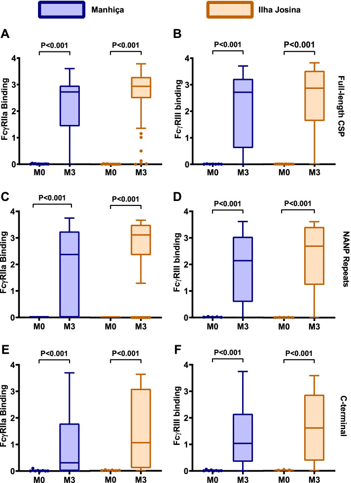
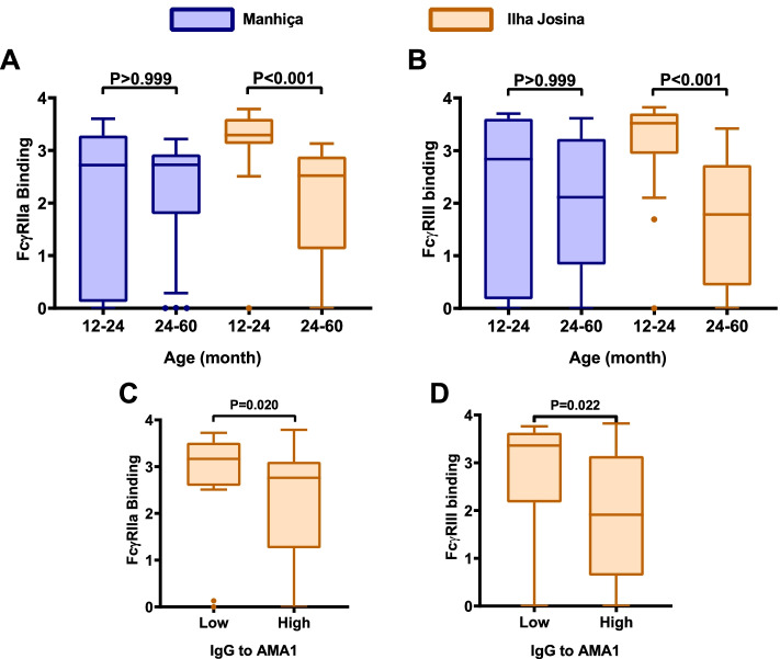
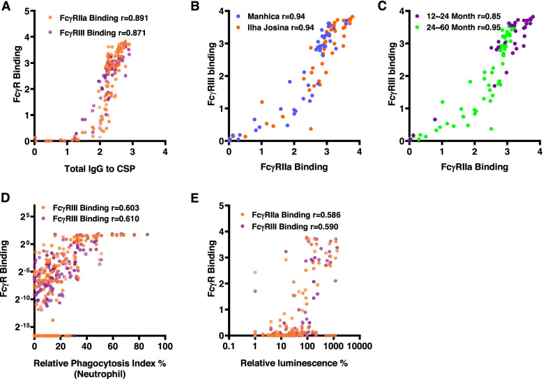
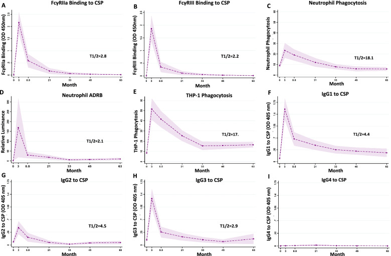
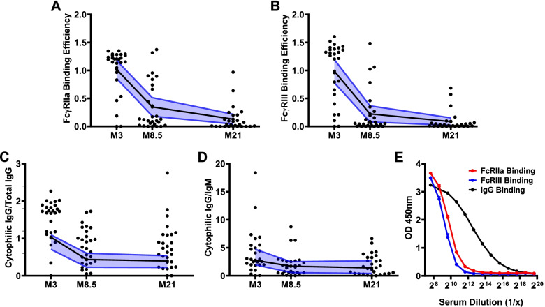
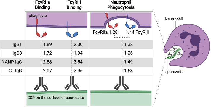
Results: RTS,S vaccination induced CSP-specific IgG with FcγRIIa and FcγRIII binding activity and promoted phagocytosis by neutrophils, THP-1 monocytes, and primary human monocytes, neutrophil ADRB activity, and NK cell activation. Responses were highly heterogenous among children, and the magnitude of neutrophil phagocytosis by antibodies was relatively modest, which may reflect modest vaccine efficacy. Induction of functional antibodies was lower among children with higher malaria exposure. Functional antibody magnitude and the functional activity of antibodies largely declined within a year post-vaccination, and decay were highest in the first 6 months, consistent with the decline in vaccine efficacy over that time. Decay rates varied for different antibody parameters and decay was slower for neutrophil phagocytosis. Biostatistical modelling suggested IgG1 and IgG3 contribute in promoting FcγR binding and phagocytosis, and IgG targeting the NANP-repeat and C-terminal regions CSP were similarly important for functional activities.

Conclusions: Results provide new insights to understand the modest and time-limited efficacy of RTS,S in children and the induction of antibody functional activities. Improving the induction and maintenance of antibodies that promote phagocytosis and cellular functions, and combating the negative effect of malaria exposure on vaccine responses are potential strategies for improving RTS,S efficacy and longevity.

Keywords: Antibodies; Children; Fcγ-receptor; Malaria; Monocytes; Neutrophils; Phagocytosis; Vaccines.


Conflict of interest statement

The authors have no conflicts of interest to declare. All co-authors have seen and agree with the contents of the manuscript and there is no financial interest to report.

Figures

Fig. 1
Fig. 1
RTS,S vaccine-induced antibodies interact with FcγRs. Serum samples from children in the RTS,S vaccine group from Manhiça (blue box plots, n=50) and Ilha Josina (orange box plots, n=49) study sites were tested for antibodies that bind FcγRIIa (left panels; A, C, E) and FcγRIII (right panels; B, D, F). Samples collected at baseline (month 0, M0) and after the third vaccination (month 3, M3) were tested against A, B full-length CSP, C, D central-repeat (NANP) and E, F C-terminal (CT) regions of CSP. Boxes represent the interquartile range (IQR) with median (bar), and whiskers represent the highest and lowest values within 1.5× IQR. Values in the Y-axis indicate the optical density (OD) at 450nm. The percentage of children with a positive response are shown in Additional file 1: Figure S1, and reactivities between paired samples were compared using Wilcoxon’s signed rank sum test
Fig. 2
Fig. 2
RTS,S vaccine-induced antibodies mediate cellular immune responses. AC Serum samples from children in the RTS,S vaccine group (Manhiça cohort) collected at baseline (month 0, M0) and after vaccination (month 3, M3) were tested for the ability to promote A opsonic phagocytosis of CSP-coated beads by neutrophils (n=30), B antibody-dependent respiratory burst (OD units) by neutrophils against CSP (n=15), C opsonic phagocytosis of CSP-coated beads by THP1 cells (n=30). D Opsonic phagocytosis by primary monocytes (n=37 (22 from Manhica, 15 from Ilha Josina cohort)). Data are shown as box plots whereby boxes represent the interquartile range (IQR) with median (bar), and whiskers represent the highest and lowest values within 1.5× IQR. The reactivities between paired samples were compared using the Wilcoxon matched-pairs signed-rank test. Phagocytosis is reported as percentage relative to a positive control (rabbit IgG to CSP), ADRB is measured as luminescence units. E Pooled samples from M0 (blue lines or bars, n=99) and M3 (red lines or bars, n=99) were tested for opsonic phagocytosis of CSP-coated beads by THP-1 cells in the presence (dashed lines) and absence (solid lines) of non-immune human serum (HS). Dots and error bars represent the mean and standard error from two independent experiments. F Pooled samples from M0 and M3 (n=99) were tested for opsonic phagocytosis by neutrophils and monocytes in a whole leukocyte assay. Data show the number of CSP-coated beads phagocytosed by 100 cells. Dots and error bars represent the mean and standard error from two independent experiments using blood from two donors. Phagocytosis was higher for neutrophils than monocytes (P=0.003, two-way repeated measures ANOVA). G The same pooled samples from M0 and M3 were tested for activation of NK cells indicating ADCC activity. Y-axis indicates the percentage of NK cells that were positive for CD107a staining by flow cytometry (P<0.001, two-way repeated measures ANOVA). Dots and error bars represent the mean and standard error based on data from 4 experiments (different NK cell donors)
Fig. 3
Fig. 3
Induction of FcγR-binding antibodies to CSP in younger and older children. Serum samples from children in the RTS,S vaccine group were stratified into younger (12–24 months) and older (24–60 months) age groups from Manhiça (blue box plots; n=11 and n=39, respectively) and Ilha Josina (orange box plots; n=24 and n=26, respectively) study sites. Samples collected after vaccination at month 3 were tested for A FcγRIIa and B FcγRIII-binding to CSP. Boxes represent the interquartile range (IQR) with median (bar) and whiskers represent the highest and lowest values within 1.5× IQR. Y-axis data represent optical density (OD) at 450nm. The percentage of children with a positive response is shown in Additional file 1: Figure S3, and reactivities between unpaired samples were compared using the Kruskal-Wallis rank sum test. For comparisons between Manhica and Ilha Josina: 12–24m age group, p=0.012 for FcγRIIa and p=0.091 for FcγRIII; for the 24–60m age group p>0.05 for both FcγRs. Results for the NANP-repeat and CT regions are shown in Additional file 1: Figure S3. Serum samples from children in the Ilha Josina study site (n=49) were also tested for IgG to the merozoite antigen AMA1 which is an established biomarker of malaria exposure (C, D). Children were classified as having low or high AMA1 IgG based on being below or above the median. Children with low AMA1 IgG had significantly higher FcγR-binding antibodies; Mann-Whitney U-test
Fig. 4
Fig. 4
Correlations between antibody response types among children vaccinated with RTS,S. Serum samples from children vaccinated with RTS,S from the Manhiça and Ilha Josina study sites at M3 were tested for FcγRIIa/III-binding to CSP and serum samples from children in the Manhiça cohort were also tested for opsonic phagocytosis and ADRB with neutrophils. Individual samples are shown as the following scatter plots: A anti-CSP IgG and FcγRIIa or FcγRIII-binding (n=100); B FcγRIIa and FcγRIII binding by study site (Manhiça, n=50 and Ilha Josina, n=49); C FcγRIIa and FcγRIII binding to CSP by age group (younger, n=35 and older, n=65); D FcγRIIa or FcγRIII-binding and opsonic phagocytosis by neutrophils; and E FcγRIIa or FcγRIII binding and ADRB. Correlations were evaluated using Spearman’s correlation coefficient (r). P-values <0.001 for all correlations. FcγR binding data are reported as optical density (OD) at 450nm
Fig. 5
Fig. 5
Kinetics of RTS,S vaccine-induced functional antibodies over time. A subset of children in the RTS,S vaccine group from the Manhiça study site was followed up over 5 years post-vaccination. Serum samples were collected at baseline (month 0, M0), 30 days after the third vaccination (month 3, M3) and later time points (M8.5, M21, M33, M45 and M63). Samples were tested for A FcγRIIa-binding and B FcγRIII-binding to CSP, C opsonic phagocytis by neutrophils (n=33) and D antibody-dependent respiratory burst (ADRB) in neutrophils and E opsonic phagocytosis by THP-1 cells. Antibodies were also tested for IgG subclasses to CSP, including F IgG1, G IgG2, H IgG3 and I IgG4. Data from immunologic assays were analysed in generalised linear mixed model (GLMM). The dashed lines represent the predicted means, the shaded area represents the 95% CIs and T1/2 (in months) indicates the estimated half-lives from the generalised linear mixed model. Half-life represents the time for magnitude to reduce by 50% from the M3 time-point. Given the very low magnitude of IgG4, it was not possible to estimate a half-life for this parameter. Further details are provided in the Additional file 1: Table S1 and S2
Fig. 6
Fig. 6
FcγR-binding efficiencies wane after RTS,S vaccination. Among children in the RTS,S vaccine group from the Manhiça study site, serum samples collected 30 days after vaccination (M3) and later time points (M8.5 and M21) were tested for various antibody responses to full-length CSP. FcγRIIa and FcγRIII binding efficiency was calculated as FcγR-binding relative to IgG magnitude for each sample at each time point. A FcγRIIa and B FcγRIII binding efficiency reduced over time (n=28, Friedman test p<0.001, respectively); C ratio of cytophilic IgG subclasses (IgG1 and IgG3) to total IgG (n=27, Friedman test p<0.001) and D ratio of cytophilic IgG subclasses to IgM (n=26, Friedman test p=0.003). Later follow-up time points were not included because the magnitude of antibodies was very low. Individual samples are shown along with the median (black lines) and 95% CI (blue lines). E Pooled samples collected at M3 (n=99) were tested at various dilutions for IgG (black line) and FcγR-binding to CSP (red line and blue line); dots and error bars represent mean and standard errors from 3 independent experiments
Fig. 7
Fig. 7
Antibody parameters correlated with functional activity. Antibodies against CSP opsonise sporozoites and interact with FcγRs to promote phagocytosis, which is predominantly mediated by neutrophils. Rate ratios are shown for the relationship between IgG subclasses and IgG to NANP-repeat and C-terminal domains of CSP, and antibody functional activities (FcγRIIa or III-binding and neutrophil phagocytosis). Rate ratios are also shown for the relationship between FcγRIIa and FcγRIII binding and neutrophil phagocytosis. Values are rate ratios and represent the percent change in participants’ antibody functional activities for each unit increase in IgG reactivity or FcγR binding. p≤0.001 for all rate ratios except neutrophil phagocytosis with IgG3 (p=0.023) and IgG to NANP (p=0.023). See Additional file 1: Tables S4 to S11 for details. Figure created with BioRender.

Similar articles

Cited by

References

    1. World Health Organization . World Malaria Report 2021. Geneva: World Health Organization; 2021.
    1. World Health Organization . Malaria vaccine: WHO position paper - January 2016. 2016.
    1. World Health Organization . Ghana, Kenya and Malawi to take part in WHO malaria vaccine pilot programme. 2017.
    1. Bejon P, Lusingu J, Olotu A, Leach A, Lievens M, Vekemans J, Mshamu S, Lang T, Gould J, Dubois MC, et al. Efficacy of RTS,S/AS01E vaccine against malaria in children 5 to 17 months of age. N Engl J Med. 2008;359(24):2521–2532. doi: 10.1056/NEJMoa0807381. - DOI - PMC - PubMed
    1. Rts SCTP, Agnandji ST, Lell B, Soulanoudjingar SS, Fernandes JF, Abossolo BP, Conzelmann C, Methogo BG, Doucka Y, Flamen A, et al. First results of phase 3 trial of RTS,S/AS01 malaria vaccine in African children. N Engl J Med. 2011;365(20):1863–1875. doi: 10.1056/NEJMoa1102287. - DOI - PubMed

Publication types

Associated data