

Regional brain volume prior to treatment is linked to outcome after cognitive rehabilitation in traumatic brain injury
- PMID: 36002956
- PMCID: PMC9421497
- DOI: 10.1016/j.nicl.2022.103126
Regional brain volume prior to treatment is linked to outcome after cognitive rehabilitation in traumatic brain injury
Abstract
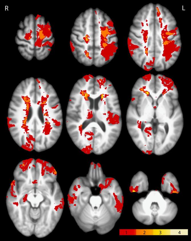
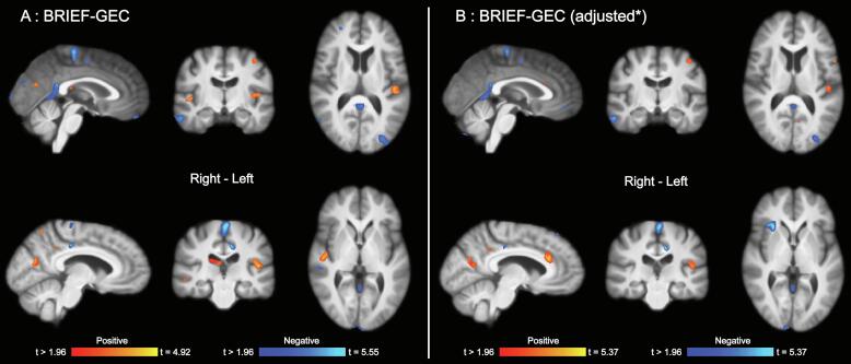
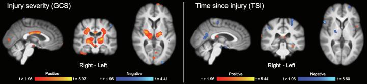
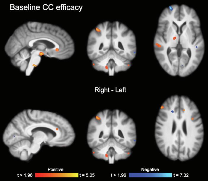
Cognitive rehabilitation is useful for many after traumatic brain injury (TBI), but we lack critical knowledge about which patients benefit the most from different approaches. Advanced neuroimaging techniques have provided important insight into brain pathology and systems plasticity after TBI, and have potential to inform new practices in cognitive rehabilitation. In this study, we aimed to identify candidate structural brain measures with relevance for rehabilitation of cognitive control (executive) function after TBI. Twenty-eight patients (9 female, mean age 40.5 (SD = 13.04) years) with TBI (>21 months since injury) that participated in a randomized controlled cognitive rehabilitation trial (NCT02692352) were included in the analyses. Regional brain volume was extracted from T1-weighted MRI scans before treatment using tensor-based morphometry. Both positive and negative associations between treatment outcome (everyday cognitive control function) and regional brain volume were observed. The most robust associations between regional brain volume and improvement in function were observed in midline fronto-parietal regions, including the anterior and posterior cingulate cortices. The study provides proof of concept and valuable insight for planning future studies focusing on neuroimaging in cognitive rehabilitation after TBI.
Keywords: Brain injury; Executive function; Magnetic resonance imaging; Personalized treatment; Rehabilitation medicine.
Copyright © 2022 The Author(s). Published by Elsevier Inc. All rights reserved.
Conflict of interest statement
The authors declare that they have no known competing financial interests or personal relationships that could have appeared to influence the work reported in this paper.
Figures
Similar articles
-
Executive function training in chronic traumatic brain injury patients: study protocol.Trials. 2019 Jul 15;20(1):435. doi: 10.1186/s13063-019-3526-x. Trials. 2019. PMID: 31307502 Free PMC article.
-
Relevance of neuroimaging for neurocognitive and behavioral outcome after pediatric traumatic brain injury.Brain Imaging Behav. 2018 Feb;12(1):29-43. doi: 10.1007/s11682-017-9673-3. Brain Imaging Behav. 2018. PMID: 28092022 Free PMC article.
-
Cognitive training benefit depends on brain injury location in adolescents with traumatic brain injury: a pilot study.Eur J Phys Rehabil Med. 2019 Oct;55(5):585-594. doi: 10.23736/S1973-9087.18.05548-X. Epub 2018 Dec 14. Eur J Phys Rehabil Med. 2019. PMID: 30547494
-
Neuroplastic Changes Induced by Cognitive Rehabilitation in Traumatic Brain Injury: A Review.Neurorehabil Neural Repair. 2017 Sep;31(9):800-813. doi: 10.1177/1545968317723748. Epub 2017 Aug 8. Neurorehabil Neural Repair. 2017. PMID: 28786307 Review.
-
Cognitive rehabilitation post traumatic brain injury: A systematic review for emerging use of virtual reality technology.J Clin Neurosci. 2019 Aug;66:209-219. doi: 10.1016/j.jocn.2019.04.026. Epub 2019 May 10. J Clin Neurosci. 2019. PMID: 31085075
References
-
- Amyot F., Arciniegas D.B., Brazaitis M.P., Curley K.C., Diaz-Arrastia R., Gandjbakhche A., Herscovitch P., Hinds S.R., Manley G.T., Pacifico A., Razumovsky A., Riley J., Salzer W., Shih R., Smirniotopoulos J.G., Stocker D. A Review of the Effectiveness of Neuroimaging Modalities for the Detection of Traumatic Brain Injury. J. Neurotrauma. 2015;32(22):1693–1721. doi: 10.1089/neu.2013.3306. - DOI - PMC - PubMed
-
- Becker F., Kirmess M., Tornås S., Løvstad M., Parente R. A description of cognitive rehabilitation at Sunnaas Rehabilitation Hospital—Balancing comprehensive holistic rehabilitation and retraining of specific functional domains. NeuroRehabilitation. 2014;34(1):87–100. - PubMed
Publication types
MeSH terms
Associated data
LinkOut - more resources
Full Text Sources
Medical
