Review
. 2022 Aug 8:21:e20210193.
doi: 10.1590/1677-5449.202101931. eCollection 2022.

Surgical access to the distal cervical segment of the internal carotid artery and to a high carotid bifurcation - integrative literature review and protocol proposal

Affiliations
Review

Surgical access to the distal cervical segment of the internal carotid artery and to a high carotid bifurcation - integrative literature review and protocol proposal

Nicolau Conte et al. J Vasc Bras. .

Abstract

Several different maneuvers have been described for obtaining access to the distal cervical segment of the internal carotid artery or to a high carotid bifurcation. However there are different approaches to systematization of these techniques. The objective of this study is to review the techniques described and propose a practical protocol to support selection of the most appropriate technique for each case. The review is based on the results of database searches on PubMed Central, the Virtual Health Library (BVSalud), and SciELO for articles on the subject published in English or Portuguese from 1980 to 2021. Among the different maneuvers described, it appears reasonable that the first two steps should be to obtain access at the sternocleidomastoid muscle, followed by section or retraction of the digastric muscle posterior belly. If needed, temporary unilateral mandibular subluxation is an additional resource that is preferable to division of the styloid apparatus process, because of its lesser potential for morbidity. Even wider exposure can be obtained using mandibular osteotomies.

Resumo: Diversas manobras já foram descritas para o acesso ao segmento distal cervical da artéria carótida interna ou à bifurcação carotídea alta; entretanto, há divergências na sistematização dessas técnicas. O objetivo deste estudo é revisar as técnicas descritas e propor um protocolo prático que auxilie na seleção da técnica mais adequada para cada caso. Para isso, foi realizada uma busca nas bases de dados PubMed Central, Biblioteca Virtual em Saúde e SciELO por artigos sobre o tema, em língua inglesa ou portuguesa, publicados entre os anos de 1980 e 2021. Entre as manobras descritas, parece razoável que as duas etapas iniciais sejam a abordagem ao músculo esternocleidomastóideo, seguida pela secção/retração do ventre posterior do músculo digástrico. Caso necessário, a subluxação mandibular temporária unilateral é um recurso adicional e preferível à divisão do aparato estiloide, devido ao menor potencial de morbidade. Exposições ainda mais amplas podem ser obtidas com as osteotomias mandibulares.

Keywords: carotid artery diseases; internal carotid artery; mandibular osteotomy; temporomandibular joint.


Conflict of interest statement

Conflicts of interest: No conflicts of interest declared concerning the publication of this article.

Figures

Figure 1
Figure 1. Flow diagram illustrating selection of articles. ICA: internal carotid artery.
Figure 2
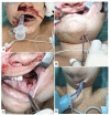
Figure 2. Submental conversion of oral endotracheal intubation (OEI). (A) Patient on OEI; (B) Submental access; (C) Dissection of the floor of the mouth; (D) Submental conversion of OEI.
Figure 3
Figure 3. Surgical approach to a traumatic cold weapon injury in left cervical zone III. (A) Cervical hematoma, cutaneous plane already sutured; (B) Access by sternocleidomastoid muscle retraction and division of the digastric muscle posterior belly, revealing the common carotid artery (CCA) and proximal segment of the internal carotid artery (ICA). The distal segment of the ICA is not exposed (broken line). Foley catheter controlling bleeding at the base of the skull; (C) Exposure of the distal segment of the ICA (dotted line), distal ICA stump is clamped.
Figure 4
Figure 4. Technique for mandibular subluxation with steel wires. (A) Positioning steel wire in mandibular interdental spaces; (B) Positioning steel wire in maxillary interdental spaces; (C) Twisting steel wires individually; (D) Twisting crossed steel wires to achieve mandibular subluxation; (E) Luxated mandibular condyle in lateral view; (F) Luxated mandibular condyle in axial view.
Figure 5
Figure 5. Application of TMS to obtain access to a contaminated stent in the internal carotid artery. (A) Infection fistularizing at the level of the surgical scar; (B) Mandibular subluxation with interdental wires; (C) Access via sternocleidomastoid muscle retraction and division of the digastric muscle posterior belly; (D) Internal carotid artery reconstructed with an autologous graft (femoral artery).
Figure 6
Figure 6. Technique for temporary mandibular subluxation (TMS) employing titanium screws. (A) Perforation of bone with a 1.6 mm burr; (B) Installation of the screws; (C) TMS with twisted crossed steel wires; (D) Application of TMS to obtain access to carotid atherosclerotic disease, employing intermaxillary molding material for additional stabilization.
Figure 7
Figure 7. Application of sternocleidomastoid muscle retraction (SCMR), division of the digastric muscle posterior belly (DDMPB), and division of the styloid apparatus (DSA) to obtain access to a pseudoaneurysm after dissection of the internal carotid artery secondary to fibromuscular dysplasia. (A) Planning the cervical incision with pre-auricular extension; (B) Surgical access with SCMR + DDMPB and exposure of the styloid process; (C) Resection of the styloid process; (D) Reconstruction of the internal carotid artery with a venous graft.
Figure 8
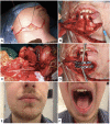
Figure 8. Application of the mandibular osteotomy technique for exeresis of carotid body tumor. (A) Cervical incision with mental extension; (B) Osteotomy of the mandibular symphysis; (C) Carotid reconstruction with a venous graft; (D) Fixation of the osteotomy with 2.0 mm titanium miniplates; (E) and (F) Patient in second postoperative month.
Figure 9
Figure 9. Images illustrating techniques for access to the distal segment of the internal carotid artery (ICA); planned using computed tomography images processed with Mimics® 24.0 software (Materialise, Leuven, Belgium). (A) Access by retraction of the sternocleidomastoid muscle (SCMR); (B) Access by SCMR and division of the digastric muscle posterior belly (DDMPB); (C) Access by SCMR + DDMPB and division of the styloid apparatus (DSA); (D) Access by SCMR + DDMPB + DSA and temporary mandibular subluxation (TMS); (E) Access by SCMR + DDMPB + DSA and mandibular osteotomy; (F) Illustration of levels of exposure of the ICA according to the technique employed.
Figure 10
Figure 10. Proposed algorithm for obtaining access to lesions of the distal segment of the internal carotid artery (DCSICA) and high carotid bifurcation (HCB). DSA: division of the styloid apparatus; SCM: sternocleidomastoid; IDS: interdental spaces; NTI: nasotracheal intubation; OEI: oral endotracheal intubation; MDO: mandibular osteotomies; R/DDMPB: retraction or division of the digastric muscle posterior belly; SM: submental; TMS: temporary mandibular subluxation; AW: airway. * Need for additional exposure of the ICA identified. ** If there is sufficient space to accommodate the oral endotracheal tube.
Figura 1
Figura 1. Fluxograma de seleção dos artigos. ACI: artéria carótida interna.
Figura 2
Figura 2. Conversão submentual da intubação orotraqueal (IOT). (A) Paciente submetido a IOT; (B) Acesso submentual; (C) Dissecção do soalho bucal; (D) Conversão submentual da IOT.
Figura 3
Figura 3. Abordagem cirúrgica de lesão traumática por arma branca em zona III cervical esquerda. (A) Hematoma cervical, plano cutâneo já suturado; (B) Acesso por retração do músculo esternocleidomastóideo e divisão do ventre posterior do músculo digástrico, evidenciando a artéria carótida comum (ACC) e o segmento proximal da artéria carótida interna (ACI). O segmento distal da ACI não está exposto (linha tracejada). Sonda de Foley tamponando o sangramento na base do crânio; (C) Exposição do segmento distal da ACI (linha pontilhada), coto distal da ACI clampeado.
Figura 4
Figura 4. Técnica de subluxação mandibular com fios de aço. (A) Posicionamento do fio de aço no espaço interdental mandibular; (B) Posicionamento do fio de aço no espaço interdental maxilar; (C) Torção dos fios de aço individualmente; (D) Torção dos fios de aço cruzados para subluxação mandibular; (E) Côndilo mandibular luxado em vista lateral; (F) Côndilo mandibular luxado em vista axial.
Figura 5
Figura 5. Aplicação da SMT para abordagem de stent contaminado na artéria carótida interna. (A) Infecção fistulizando ao nível da cicatriz cirúrgica; (B) Subluxação mandibular com amarrias interdentais; (C) Acesso por retração do músculo esternocleidomastóideo e divisão do ventre posterior do músculo digástrico; (D) Artéria carótida interna reconstruída com enxerto autólogo (artéria femoral).
Figura 6
Figura 6. Técnica de subluxação mandibular temporária (SMT) utilizando parafusos de titânio. (A) Perfuração óssea com fresa 1,6 mm; (B) Instalação dos parafusos; (C) SMT com torção dos fios de aço cruzados; (D) Aplicação da SMT para abordagem de doença carotídea aterosclerótica, com auxílio de material de moldagem intermaxilar para estabilização adicional.
Figura 7
Figura 7. Aplicação das técnicas de retração do músculo esternocleidomastóideo (RECM), divisão do ventre posterior do músculo digástrico (DVPD) e divisão do aparato estiloide (DAE) para abordagem de pseudoaneurisma após dissecção de artéria carótida interna por displasia fibromuscular. (A) Planejamento da incisão cervical com extensão pré-auricular; (B) Acesso cirúrgico RECM + DVDP e exposição do processo estiloide; (C) Ressecção do processo estiloide; (D) Reconstrução da artéria CI com enxerto venoso.
Figura 8
Figura 8. Aplicação da técnica de osteotomia mandibular para exérese de tumor de corpo carotídeo. (A) Incisão cervical com extensão mentual; (B) Osteotomia realizada na sínfise mandibular; (C) Reconstrução carotídea com enxerto venoso; (D) Fixação da osteotomia com miniplacas de titânio de 2,0 mm. (E) e (F) Paciente no segundo mês de pós-operatório.
Figura 9
Figura 9. Representação das técnicas de acesso ao segmento distal da artéria carótida interna (ACI); planejamento por imagens de tomografia computadorizada processadas no software Mimics® 24.0 (Materialise, Leuven, Belgium). (A) Acesso por retração do músculo esternocleidomastóideo (RECM); (B) Acesso por RECM e divisão do ventre posterior do músculo digástrico (DVPD); (C) Acesso por RECM + DVPD e divisão do aparato estiloide (DAE); (D) Acesso por RECM + DVPD + DAE e subluxação mandibular temporária (SMT); (E) Acesso RECM + DVPD + DAE e osteotomia mandibular; (F) Demonstração do nível de exposição da ACI de acordo com a técnica.
Figura 10
Figura 10. Algoritmo proposto para abordagem das lesões em segmento distal da artéria carótida interna (SDACI) e bifurcação carótida alta (BCA). DAE: divisão do aparato estiloide; ECM: esternocleidomastóideo; EI: espaços interdentais; INT: intubação nasotraqueal; IOT: intubação orotraqueal; OMD: osteotomias mandibulares; R/DVPD: retração ou divisão do ventre posterior do músculo digástrico; SM: submentual; SMT: subluxação mandibular temporária; VA: via aérea. * Identificou necessidade de exposição adicional da ACI. ** Quando houver espaço suficiente para acomodar o tubo orotraqueal.

References

    1. Reva VA, Pronchenko AA, Samokhvalov IM. Operative management of penetrating carotid artery injuries. Eur J Vasc Endovasc Surg. 2011;42(1):16–20. doi: 10.1016/j.ejvs.2011.01.025. - DOI - PubMed
    1. Lee TS, Ducic Y, Gordin E, Stroman D. Management of carotid artery trauma. Craniomaxillofac Trauma Reconstr. 2014;7(3):175–189. doi: 10.1055/s-0034-1372521. - DOI - PMC - PubMed
    1. Fisher DF, Jr, Clagett GP, Parker JI, et al. Mandibular subluxation for high carotid exposure. J Vasc Surg. 1984;1(6):727–733. doi: 10.1016/0741-5214(84)90002-8. - DOI - PubMed
    1. Awasthi D, Smith R. Distal cervical internal carotid artery stenosis. Skull Base Surg. 1994;4(2):110–115. doi: 10.1055/s-2008-1058980. - DOI - PMC - PubMed
    1. Beretta F, Hemida SA, Andaluz N, Zuccarello M, Keller JT. Exposure of the cervical internal carotid artery: surgical steps to the cranial base and morphometric study. Neurosurgery. 2006;59(1) Suppl 1:ONS25–34, discussion ONS25-34. - PubMed

LinkOut - more resources