

MRI appearance of chronic subdural hematoma
- PMID: 36003292
- PMCID: PMC9393587
- DOI: 10.3389/fneur.2022.872664
MRI appearance of chronic subdural hematoma
Abstract
Objective: We aimed to describe the magnetic resonance imaging (MRI) characteristics of chronic subdural hematoma (cSDH) and to ascribe MRI patterns.
Methods: A total of 20 patients having 27 subdural hematomas underwent contrast-enhanced (CE) MRI of the brain at our institution between April 2019 and May 2021. The images were independently evaluated by two experienced neuroradiologists with regard to imaging characteristics on T1w, T2w, T2*-GRE, FLAIR, diffusion-weighted magnetic resonance imaging (DWI), and CE images.
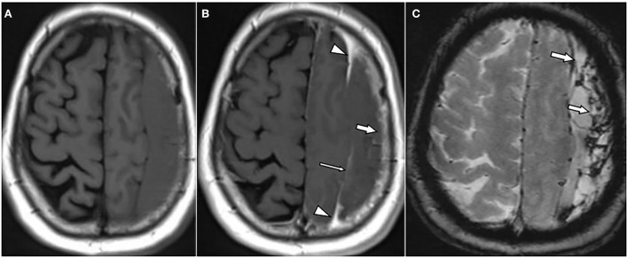
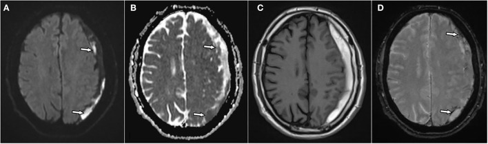
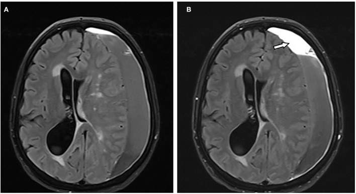
Results: The signal characteristics of cSDH on T1- and T2-weighted images were rather heterogeneous. The majority of hematomas (74%) had internal septations. Surprisingly, contrast enhancement along the outer membrane adjacent to the cranium was noticed in all hematomas. There was also contrast enhancement along the inner membrane adjacent to the brain in more than one-third of the hematomas (37%). In approximately two-thirds of the cSDH (62%), there was a mass-like enhancement of the hematoma. Most hematomas (89%) were partially hypointense on T2*-GRE and/or susceptibility-weighted imaging (SWI). Restricted diffusion was detected in approximately one-third of the hematomas (33%).
Conclusion: Consistent contrast enhancement along the outer membrane, triangular-shaped contrast enhancement at the borders of the cSDH, and infrequent enhancement of the inner membrane may help to distinguish cSDH from other entities such as empyema and tumors. Mass-like enhancement may refer to non-solid hematomas and could be an indicator for hematoma growth and a possible surrogate for successful endovascular embolization. Restricted diffusion in a subdural mass is not specific for empyema but may also be found in cSDH.
Keywords: chronic; empyema; hematoma; magnetic resonance imaging; subdural.
Copyright © 2022 Hasan, Nikoubashman, Pjontek, Stockero, Hamou and Wiesmann.
Conflict of interest statement
The authors declare that the research was conducted in the absence of any commercial or financial relationships that could be construed as a potential conflict of interest.
Figures

References
-
- Stanišić M, Hald J, Rasmussen IA, Pripp AH, Ivanović J, Kolstad F, et al. . Volume and densities of chronic subdural haematoma obtained from CT imaging as predictors of postoperative recurrence: a prospective study of 107 operated patients. Acta Neurochir. (2013) 155:323–33; discussion 33. 10.1007/s00701-012-1565-0 - DOI - PMC - PubMed
-
- Guénot M. [Chronic subdural hematoma: diagnostic imaging studies]. Neurochirurgie. (2001) 47:473–8. - PubMed
-
- Senturk S, Guzel A, Bilici A, Takmaz I, Guzel E, Aluclu MU, et al. . CT and MR imaging of chronic subdural hematomas: a comparative study. Swiss Med Wkly. (2010) 140:335–40. - PubMed
LinkOut - more resources
Full Text Sources
Miscellaneous