. 2022 Aug 19:10:e13844.
doi: 10.7717/peerj.13844. eCollection 2022.

CCL25/CCR9 interaction promotes the malignant behavior of salivary adenoid cystic carcinoma via the PI3K/AKT signaling pathway

Affiliations

CCL25/CCR9 interaction promotes the malignant behavior of salivary adenoid cystic carcinoma via the PI3K/AKT signaling pathway

Songling Chai et al. PeerJ. .

Abstract

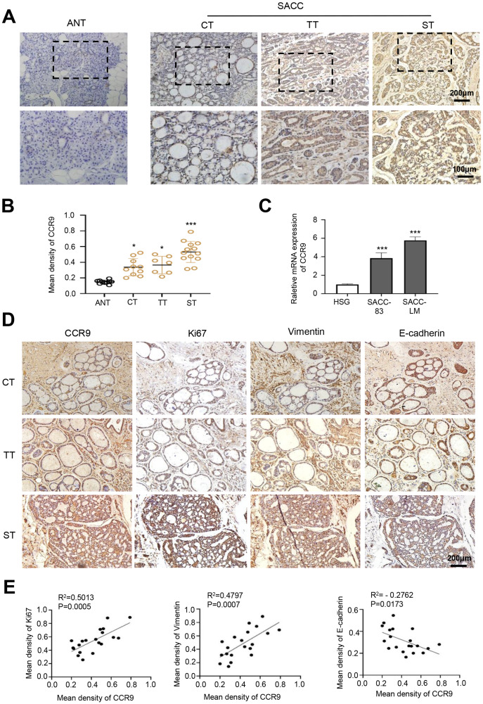
Background: CC chemokine receptor 9 (CCR9), an organ-specific chemokine receptor, interacts with its exclusive ligand CCL25 to promote tumor proliferation and metastasis. However, the effect of CCR9 on salivary adenoid cystic carcinoma (SACC) malignant behavior remains unknown. This study aimed to investigate the specific molecular mechanism by which CCR9/CCL25 modulates malignant progression in SACC.

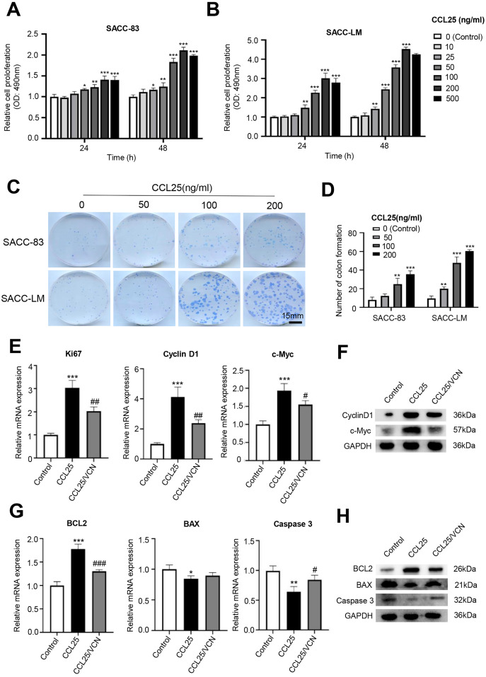
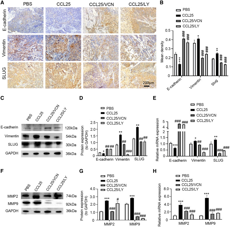
Methods: Immunohistochemistry staining and RT-qPCR analyses were performed to detect the correlation of CCR9 expression and tumor progression-associated markers in SACC. In vitro, SACC cell proliferation and apoptosis were evaluated using Cell Counting Kit-8 and colon formation, and cell migration and invasion were detected by wound healing and transwell assays. Vercirnon was used as an inhibitor of CCR9, and LY294002 was used as an inhibitor of the PI3K/AKT pathway in this study. Western blot and RT-qPCR assays were carried out to measure the downstream factors of the interaction of CCL25 and CCR9. The effect of CCL25 on the development of SACC in vivo was examined by a xenograft tumor model in nude mice following CCL25, Vercirnon and LY294002 treatment.

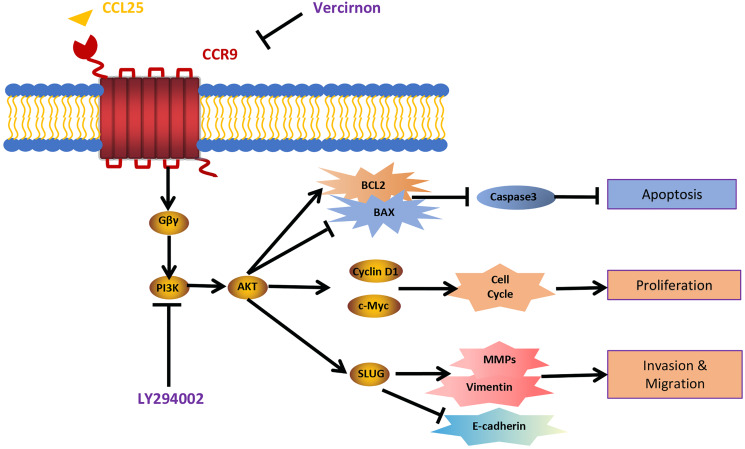
Results: CCR9 was highly expressed in SACC compared with adjacent salivary gland tissues, and its level was associated with tumor proliferation and metastases. CCL25 enhanced cell proliferation, migration, and invasion through its interaction with CCR9 and exerted an antiapoptotic effect on SACC cells. Targeting CCR9 via Vercirnon significantly reduced the phosphorylation level of AKT induced by CCL25. CCL25/CCR9 could activate its downstream factors through the PI3K/AKT signaling pathway, such as cyclin D1, BCL2 and SLUG, thus promoting SACC cell proliferation, antiapoptosis, invasion and metastasis. The in vivo data from the xenograft mouse models further proved that CCL25 administration promoted malignant tumor progression by activating the PI3K/AKT pathway.

Conclusion: The interaction of CCL25 and CCR9 promotes tumor growth and metastasis in SACC by activating the PI3K/AKT signaling pathway, offering a promising strategy for SACC treatment.

Keywords: CCL25; CCR9; Metastasis; PI3K/Akt; Proliferation; Salivary adenoid cystic carcinoma.


Conflict of interest statement

The authors declare that they have no competing interests.

Figures

Figure 1
Figure 1. Expression of CCR9 in SACC and its correlation with tumor proliferation and metastasis.
(A) Representative images showing the expression of CCR9 in different types of SACC (cribriform type, CT, n = 10; tubular type, TT, n = 7; solid type, ST, n = 13) by IHC. Adjacent normal tissue (ANT, n = 10) was used as the control group. The boxed areas in the upper panels are shown at higher magnification in the lower panels. (B) The relative fold change in CCR9 expression (A) in SACC was quantitatively analyzed (unpaired two-tailed t-test, *P < 0.05, ***P < 0.001). (C) The mRNA expression of CCR9 was determined by RT–qPCR in the SACC-83, SACC-LM and HSG cell lines (n = 3, unpaired two-tailed t-test, ***P < 0.001). (D) Representative images of the expression of tumor proliferation and invasion markers (Ki67, vimentin, and E-cadherin) by IHC in the three types of SACC. (E) Pearson correlation analysis was used to analyze the expression of Ki67, vimentin, E-cadherin and CCR9 in SACC (n = 20, two‑tailed Pearson’s correlation). Data are presented as the mean ± SD.
Figure 2
Figure 2. CCL25/CCR9 promotes the proliferation of SACC cells.
(A, B) The proliferation of SACC-83 (A) and SACC-LM (B) cells treated with different concentrations of CCL25 was tested by a CCK-8 kit at 24 h and 48 h (n = 3, unpaired two-tailed t-test; *P < 0.05, **P < 0.01, ***P < 0.001). (C) Colony-forming assay was performed to detect the proliferation of SACC-83 and SACC-LM cells treated with CCL25 (n = 3, unpaired two-tailed t-test; **P < 0.01, ***P < 0.001). (D) Quantitative analysis of the colon level of (C). (E, F) SACC-LM cells were treated with 200 ng/ml CCL25 and 10 nM CCR9 inhibitor (Vercirnon, VCN) for 48 h. The mRNA and protein expression of proliferation-related factors (Ki67, cyclin D1, and c-Myc) was detected by RT–qPCR (E) and WB (F) (n = 3, one-way ANOVA followed by Tukey’s multiple comparisons; * vs Control, ***P < 0.001; # vs CCL25, #P < 0.05, ##P < 0.01.). (G, H) The mRNA and protein expression of apoptosis-related factors (BCL2, BAX, and caspase 3) was detected by RT–qPCR (G) and WB (H) (n = 3, one-way ANOVA followed by Tukey’s multiple comparisons; * vs Control, *P < 0.05, **P < 0.01, ***P < 0.001; # vs CCL25, #P < 0.05, ###P < 0.001). Data are presented as the mean ± SD.
Figure 3
Figure 3. CCL25/CCR9 promotes SACC-LM cell migration and invasion.
(A) After SACC-LM cells were treated with different concentrations of CCL25 (0, 100, 200 ng/ml for 24 h, wound healing assays were used to test the horizontal migration ability of SACC-LM cells. (B) The wound closure area of cells after CCL25 administration was quantitatively analyzed (n = 3, unpaired two-tailed t-test; **P < 0.01, ***P < 0.001). (C) The migration and invasion abilities were detected by Transwell assays after SACC-LM cells were treated with different concentrations of CCL25 (0, 100, 200 ng/ml) for 24 h. (D) Quantitative analysis of the number of migrated and invasive cells in (C) (n = 3, unpaired two-tailed t-test; *P < 0.05, ***P < 0.001). (E, F) The mRNA and protein expression of vimentin, E-cadherin, MMP2 and MMP9 was measured by RT–qPCR (E) and WB (F). GAPDH was used as a standard control (n = 3, one-way ANOVA followed by Tukey’s multiple comparisons; * vs Control, ***P < 0.001; # vs CCL25, #P < 0.05, ###P < 0.001). Data are presented as the mean ± SD.
Figure 4
Figure 4. CCL25/CCR9 activates the PI3K/AKT signaling pathway in SACC-LM cells.
(A) WB was used to detect the activation of signaling pathways, including p-ERK1/2 (ERK1: T202/Y204; ERK2: T185/Y187), p-AKT (Ser473) p-STAT3 (Tyr705) after SACC-LM cells were treated with/without 10 nM Vercirnon for 48 h followed by 200 ng/ml CCL25 pretreatment. (B) Quantitative analysis of the protein level of p-AKT, p-ERK1/2 and p-STAT3 (n = 3, one-way ANOVA followed by Tukey’s multiple comparisons; ***P < 0.001; # vs CCL25, ##P < 0.01). Data are presented as the mean ± SD.
Figure 5
Figure 5. CCL25/CCR9 interaction enhances the proliferation and anti-apoptosis of SACC-LM cells through the PI3K/AKT pathway.
(A, B) After treatment with 200 ng/ml CCL25, different concentrations of PI3K/AKT inhibitor (LY294002, LY) were added to 5 × 10³ SACC-LM cells for 24 or 48 h. The proliferation of SACC-LM cells was detected by CCK-8 assay (n = 3, one-way ANOVA followed by Tukey’s multiple comparisons; * vs Control, ***P < 0.001; # vs CCL25, #P < 0.05, ##P < 0.01, ###P < 0.001). (C) Cells viability was detected by Live and Dead Cell Double Staining experiment. Red light represented Dead cells and green light represented living cells. (D) Quantitative analysis of the fluorescence number of dead cells per unit area. (n = 3, one-way ANOVA followed by Tukey’s multiple comparisons; * vs Control, ***P < 0.001; # vs CCL25, ###P < 0.001). (E, F) A total of 1 × 10⁵ SACC-LM cells were administered with 200 ng/ml CCL25, 10 nM LY294002 and 10 nM Vercirnon for 48 h. Proliferation-related factors (Ki67, cyclin D1, and c-Myc) were detected by RT–qPCR (E) and WB (F) (n = 3, one-way ANOVA followed by Tukey’s multiple comparisons; * vs Control, ***P < 0.001; # vs CCL25, ##P < 0.01, ###P < 0.001). (G, H) The mRNA and protein expression of apoptosis-related factors (BCL2, BAX, and caspase 3) was detected by RT–qPCR (G) and WB (H) (n = 3, one-way ANOVA followed by Tukey’s multiple comparisons; * vs Control, *P < 0.05, **P < 0.01; # vs CCL25, #P < 0.05). Data are presented as the mean ± SD.
Figure 6
Figure 6. CCL25/CCR9 interaction enhances the migration and invasion of SACC-LM cells through the PI3K/AKT pathway.
(A–E) A total of 1 × 10⁵ SACC-LM cells were administered 200 ng/ml CCL25, 10 nM PI3K/AKT inhibitor (LY294002) and 10 nM Vercirnon (VCN) for 48 h. The mRNA and protein expression of invasion- and metastasis-related proteins (vimentin, E-cadherin, MMP2 and MMP9) was detected by RT–qPCR (A–D) and WB (E) (n = 3, one-way ANOVA followed by Tukey’s multiple comparisons; * vs Control, ***P < 0.001; # vs CCL25, #P < 0.05, ###P < 0.001). (F–I) The mRNA and protein expression of EMT-related transcription factors (SLUG, SNAIL1, and TWIST) was detected by RT–qPCR (F–H) and WB (I) (n = 3, one-way ANOVA followed by Tukey’s multiple comparisons; * vs Control, **P < 0.01, ***P < 0.001; # vs CCL25, ###P < 0.001). Data are presented as the mean ± SD.
Figure 7
Figure 7. CCL25/CCR9 promotes tumor growth in a SACC xenograft mouse model by activating the PI3K/AKT signaling pathway.
(A) Schematic representation of the animal model establishment. For xenograft models, 1 × 10⁷ SACC-LM cells in 100 μl of PBS were subcutaneously injected into the right flanks of mice. After 7 days of tumor cell injection, the mice were randomly divided into four groups: PBS group, CCL25 group, CCL25/VCN group and CCL25/LY group. According to the time axis, mice were administered CCL25 on Day 7 and then given CCR9 inhibitor (Vercirnon) or PI3K/AKT inhibitor (LY294002) on Days 7 and 14, respectively, around the transplanted tumor. Then, the mice were euthanized at Day 28, and the xenograft tumors were harvested. (B) The growth curve of xenograft tumor size in nude mice was measured every three days (n = 5, one-way ANOVA followed by Tukey’s multiple comparisons; * vs Control, ***P < 0.001; # vs CCL25, ###P < 0.001). (C) Comparison of tumor tissue weights in the four groups (n = 5, one-way ANOVA followed by Tukey’s multiple comparisons; * vs Control, ***P < 0.001; # vs CCL25, ###P < 0.001). (D) Representative images from the histological staining of HE, TUNEL and Ki67 in tumor tissues. (E) Quantitative analysis of TUNEL and Ki67 staining (n = 5, one-way ANOVA followed by Tukey’s multiple comparisons; * vs Control, *P < 0.05, **P < 0.01; # vs CCL25, ##P < 0.01, ###P < 0.001). (F) Proliferation- and apoptosis-related transcription factors were detected in tumor tissues by WB. (G) Quantitative analysis of protein expression in (F) (n = 5, one-way ANOVA followed by Tukey’s multiple comparisons; * vs Control, ***P < 0.001; # vs CCL25, ###P < 0.001). (H) mRNA expression of proliferation- and apoptosis-related factors was measured by RT–qPCR (n = 3, one-way ANOVA followed by Tukey’s multiple comparisons; * vs Control, **P < 0.01, ***P < 0.001; # vs CCL25, ###P < 0.001). Data are presented as the mean ± SD.
Figure 8
Figure 8. CCL25/CCR9 promotes invasion and migration in a SACC xenograft mouse model by activating the PI3K/AKT signaling pathway.
(A) The expression of tumor invasion- and metastasis-related factors (E-cadherin, vimentin and SLUG) in xenograft tumor tissues was detected by IHC staining. (B) The expression level was analyzed quantitatively (n = 5, one-way ANOVA followed by Tukey’s multiple comparisons; * vs Control, *P < 0.05, **P < 0.01, ***P < 0.001; # vs CCL25, ###P < 0.001). (C, D) The protein expression of E-cadherin, vimentin and SLUG in xenograft tumors was measured by WB and statistically analyzed (n = 5, one-way ANOVA followed by Tukey’s multiple comparisons; * vs Control, *P < 0.05, **P < 0.01; # vs CCL25, ##P < 0.01, ###P < 0.001). (E) The mRNA expression of E-cadherin, vimentin and SLUG in xenograft tumors was assessed by RT–qPCR (n = 3, one-way ANOVA followed by Tukey’s multiple comparisons; * vs Control, *P < 0.05, **P < 0.01, ***P < 0.001; # vs CCL25, ##P < 0.01, ###P < 0.001). (F, G) The protein expression of ECM-related proteins (MMP2 and MMP9) in xenograft tumors and statistical analysis (n = 5, one-way ANOVA followed by Tukey’s multiple comparisons; * vs Control, *P < 0.05, ***P < 0.001; # vs CCL25, ###P < 0.001). (H) The mRNA expression levels of MMP2 and MMP9 were measured by RT–qPCR (n = 3, one-way ANOVA followed by Tukey’s multiple comparisons; * vs Control, ***P < 0.001; # vs CCL25, ###P < 0.001). Data are presented as the mean ± SD.
Figure 9
Figure 9. Schematic model illustrating the PI3K/AKT pathway associated with CCRL25/CCR9-induced malignant biological behavior in SACC.
CCL25 acts on CCR9, a G protein-coupled receptor located on the SACC cell membrane, resulting in activation of the G protein and release of the Gβγ subunit, thereby activating the PI3K/AKT signaling pathway. Vercirnon is a CCR9 inhibitor, and LY294002 is a blocker of the PI3K/AKT pathway. PI3K/AKT pathway activation can upregulate the mitochondrial-mediated anti-apoptotic factor BCL2, thus inhibiting caspase 3-dependent tumor cell apoptosis. Furthermore, CCL25 activation of the PI3K/AKT pathway leads to an increase in the expression of the cell cycle regulatory protein cyclin D1 and the protooncogene c-Myc, thus promoting the proliferation of tumor cells. Furthermore, the activation of the PI3K/AKT pathway activates its downstream transcription factor SLUG, leading to regulation of the expression of EMT-related factors (E-cadherin and vimentin) and ECM degradation-related factors (MMPs), which enhances the migration and invasion abilities of SACC cells.

Similar articles

Cited by

References

    1. Atanes P, Lee V, Huang GC, Persaud SJ. The role of the CCL25-CCR9 axis in beta-cell function: potential for therapeutic intervention in type 2 diabetes. Metabolism. 2020;113:154394. doi: 10.1016/j.metabol.2020.154394. - DOI - PubMed
    1. Bai JW, Chen MN, Wei XL, Li YC, Lin HY, Chen M, Li JW, Du CW, Man K, Zhang GJ. The zinc-finger transcriptional factor Slug transcriptionally downregulates ERα by recruiting lysine-specific demethylase 1 in human breast cancer. Oncogenesis. 2017;6(5):e330. doi: 10.1038/oncsis.2017.38. - DOI - PMC - PubMed
    1. Bai J, Li L, Kou N, Bai Y, Zhang Y, Lu Y, Gao L, Wang F. Low level laser therapy promotes bone regeneration by coupling angiogenesis and osteogenesis. Stem Cell Research & Therapy. 2021;12(1):432. doi: 10.1186/s13287-021-02493-5. - DOI - PMC - PubMed
    1. Brabletz T, Kalluri R, Nieto MA, Weinberg RA. EMT in cancer. Nature Reviews Cancer. 2018;18(2):128–134. doi: 10.1038/nrc.2017.118. - DOI - PubMed
    1. Chau NG, Hotte SJ, Chen EX, Chin SF, Turner S, Wang L, Siu LL. A phase II study of sunitinib in recurrent and/or metastatic adenoid cystic carcinoma (ACC) of the salivary glands: current progress and challenges in evaluating molecularly targeted agents in ACC. Annals of Oncology. 2012;23(6):1562–1570. doi: 10.1093/annonc/mdr522. - DOI - PubMed

Publication types