. 2022 Aug 8:13:933381.
doi: 10.3389/fgene.2022.933381. eCollection 2022.

Functional Evaluation and Genetic Landscape of Children and Young Adults Referred for Assessment of Bronchiectasis

Affiliations

Functional Evaluation and Genetic Landscape of Children and Young Adults Referred for Assessment of Bronchiectasis

Jeffrey Fong Ting Chau et al. Front Genet. .

Abstract

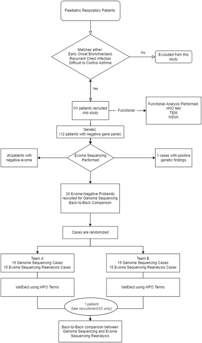
Bronchiectasis is the abnormal dilation of the airway which may be caused by various etiologies in children. Beyond the more recognized cause of bacterial and viral infections and primary immunodeficiencies, other genetic conditions such as cystic fibrosis and primary ciliary dyskinesia (PCD) can also contribute to the disease. Currently, there is still debate on whether genome sequencing (GS) or exome sequencing reanalysis (rES) would be beneficial if the initial targeted testing results returned negative. This study aims to provide a back-to-back comparison between rES and GS to explore the best integrated approach for the functional and genetics evaluation for patients referred for assessment of bronchiectasis. In phase 1, an initial 60 patients were analyzed by exome sequencing (ES) with one additional individual recruited later as an affected sibling for ES. Functional evaluation of the nasal nitric oxide test, transmission electron microscopy, and high-speed video microscopy were also conducted when possible. In phase 2, GS was performed on 30 selected cases with trio samples available. To provide a back-to-back comparison, two teams of genome analysts were alternatively allocated to GS or rES and were blinded to each other's analysis. The time for bioinformatics, analysis, and diagnostic utility was recorded for evaluation. ES revealed five positive diagnoses (5/60, 8.3%) in phase 1, and four additional diagnoses were made by rES and GS (4/30, 13%) during phase 2. Subsequently, one additional positive diagnosis was identified in a sibling by ES and an overall diagnostic yield of 10/61 (16.4%) was reached. Among those patients with a clinical suspicion of PCD (n = 31/61), the diagnostic yield was 26% (n = 8/31). While GS did not increase the diagnostic yield, we showed that a variant of uncertain significance could only be detected by GS due to improved coverage over ES and hence is a potential benefit for GS in the future. We show that genetic testing is an essential component for the diagnosis of early-onset bronchiectasis and is most effective when used in combination with functional tools such as TEM or HSVM. Our comparison of rES vs. GS suggests that rES and GS are comparable in clinical diagnosis.

Keywords: early-onset bronchiectasis; exome sequencing; genome sequencing; high-speed video microscopy; primary ciliary dyskinesia; transmission electron microscopy.


Conflict of interest statement

The authors declare that the research was conducted in the absence of any commercial or financial relationships that could be construed as a potential conflict of interest.

Figures

FIGURE 1
FIGURE 1
Diagnostic workflow. The diagram shows this study design and workflow. In phase 1, 61 patients were recruited into the study for genetic and functional analysis. In phase 2, 30 exome-negative probands were recruited for GS and rES back-to-back comparison analysis. HSVM = high-speed video microscopy; HPO = human phenotype ontology; nNO test = nasal NO test; TEM = transmission electron microscopy.
FIGURE 2
FIGURE 2
Transmission electron microscopy results of suspected early-onset bronchiectasis patients in Hong Kong. (A) Case 12—Confirmed CF case showed a normal ciliary ultrastructure. (B) Case 5—A homozygous recessive DNAAF3 c 493G>C variant was identified. TEM results showed an outer and inner dynein arm (arrows) defect which is similar to previously reported cases (C). Case 25 cilia proximal arm—A compound heterozygous variants could be seen in DNAH9. The literature has shown that subtle outer dynein arm defect can be seen in TEM. The ciliary ultrastructure analysis in the proximal arm shows a normal ultrastructure (D). Case 25 cilia distal arm shows a subtle outer dynein arm (arrow) defect. (E) Case 63—TEM results showed a normal ciliary ultrastructure. A pathogenic homozygous recessive DNAH11 c.11749_11752delGTTA was identified and confirmed the clinical diagnosis despite the normal ultrastructure. HSVM showed a static beat pattern in 50% of cilia samples (F). Case 6—TEM results showed that 47% of cilia were in a disarranged pattern (arrows). This matches the literature of loss-of-function mutations in the CCDC40 gene. The frameshift deletion detected was CCDC40 c.626_627del. (G). Case 36—A trio-based ES revealed a frameshift deletion mutation on the RSPH4A gene, c.1774_1775del. Two separate TEM results showed 43 and 35% of ciliary samples with missing/extra-central microtubules (arrows). Previously reported literature has shown that patients with RSPH4A mutations are also presented with circular beating patterns and cilia without central microtubules (H). Case 2—A compound heterozygous of two variants detected in trans within the DNAI1 gene was seen in both trio-based rES and GS. ODA (arrows) defect was also detected in the TEM investigation which showed an average of 3.4 outer dynein arms detected per cilia (arrowheads, inner dynein arm). (bar = 100 nm).
FIGURE 3
FIGURE 3
Nucleotide by nucleotide coverage of DNAAF3 in ES and GS. The line plot shows the average coverage of each nucleotide in the DNAAF3 gene. (A) Coverage of ES split across library preparation kits. Across 30 ES samples that used Truseq, an average of 0× read depth was seen for exon 5 of DNAAF3. (B) Coverage of GS across the DNAAF3 gene.

Similar articles

Cited by

References

    1. Abou Tayoun A. N., Pesaran T., Distefano M. T., Oza A., Rehm H. L., Biesecker L. G., et al. (2018). Recommendations for Interpreting the Loss of Function PVS1 ACMG/AMP Variant Criterion. Hum. Mutat. 39, 1517–1524. 10.1002/humu.23626 - DOI - PMC - PubMed
    1. Abyzov A., Urban A. E., Snyder M., Gerstein M. (2011). CNVnator: an Approach to Discover, Genotype, and Characterize Typical and Atypical CNVs from Family and Population Genome Sequencing. Genome Res. 21, 974–984. 10.1101/gr.114876.110 - DOI - PMC - PubMed
    1. Afzelius B. A., Stenram U. (2006). Prevalence and Genetics of Immotile-Cilia Syndrome and Left-Handedness. Int. J. Dev. Biol. 50, 571–573. 10.1387/ijdb.052132ba - DOI - PubMed
    1. Boon M., Wallmeier J., Ma L., Loges N. T., Jaspers M., Olbrich H., et al. (2014). MCIDAS Mutations Result in a Mucociliary Clearance Disorder with Reduced Generation of Multiple Motile Cilia. Nat. Commun. 5, 4418. 10.1038/ncomms5418 - DOI - PubMed
    1. Chen X., Schulz-Trieglaff O., Shaw R., Barnes B., Schlesinger F., Källberg M., et al. (2016). Manta: Rapid Detection of Structural Variants and Indels for Germline and Cancer Sequencing Applications. Bioinformatics 32, 1220–1222. 10.1093/bioinformatics/btv710 - DOI - PubMed

LinkOut - more resources