Review
. 2022 Aug 8:12:945376.
doi: 10.3389/fonc.2022.945376. eCollection 2022.

Small extracellular vesicle DNA-mediated horizontal gene transfer as a driving force for tumor evolution: Facts and riddles

Affiliations
Review

Small extracellular vesicle DNA-mediated horizontal gene transfer as a driving force for tumor evolution: Facts and riddles

Gábor Valcz et al. Front Oncol. .

Abstract

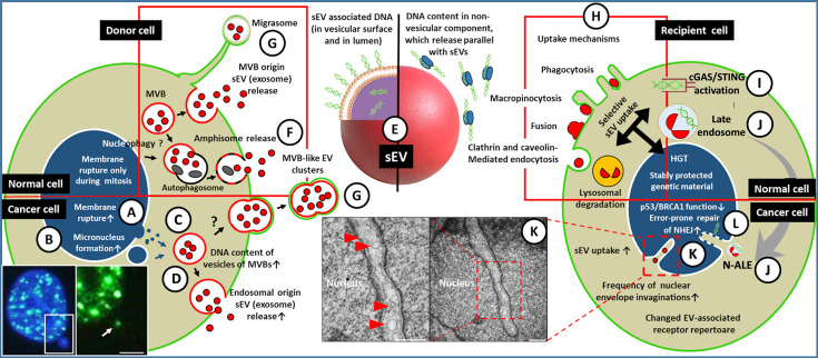
The basis of the conventional gene-centric view on tumor evolution is that vertically inherited mutations largely define the properties of tumor cells. In recent years, however, accumulating evidence shows that both the tumor cells and their microenvironment may acquire external, non-vertically inherited genetic properties via horizontal gene transfer (HGT), particularly through small extracellular vesicles (sEVs). Many phases of sEV-mediated HGT have been described, such as DNA packaging into small vesicles, their release, uptake by recipient cells, and incorporation of sEV-DNA into the recipient genome to modify the phenotype and properties of cells. Recent techniques in sEV separation, genome sequencing and editing, as well as the identification of new secretion mechanisms, shed light on a number of additional details of this phenomenon. Here, we discuss the key features of this form of gene transfer and make an attempt to draw relevant conclusions on the contribution of HGT to tumor evolution.

Keywords: cell-cell communication; exosomes; extracellular vesicles; horizontal gene transfer; tumor evolution.


Conflict of interest statement

The authors declare that the research was conducted in the absence of any commercial or financial relationships that could be construed as a potential conflict of interest.

Figures

Figure 1
Figure 1
A simplified representation of sEV-mediated HGT among tumor/microenvironmental cells. (A) The nuclear gDNA is discharged to the cytoplasmic region of the EV releasing cell (left) through the rupture of the nuclear envelope or by the formation of micronucleus (the fluorescent microscopic image shows a double-strand break (DSB) site in the micronucleus of HT-29 cell [white arrow, γH2AX staining, scale bar: 2 µm)] (B). (C) From the cytoplasm or from micronuclei the gDNA translocates into the intraluminal vesicles (future exosomes) of multivesicular bodies. (D) Both the gDNA content of the exosomes and their release are increased in tumor cells, especially upon the effect of therapeutic stress. (E) The gDNA may be transferred either in the lumen of sEVs and/or on the exofacial EV surface, or independently as a non-vesicular component. The gDNA content of sEVs might depend on their origin, like exocytosis of MVBs (D), amphisomes (F), or sEV discharge from en bloc released MVB-like EV clusters or migrasomes (G). The red frame indicates that all listed processes (i.e., migrasome formation, exosome secretion, amphisome exocytosis) may occur in both normal and tumor cells. (H) Uptake of the released sEVs by a recipient cell (right) may include receptor-mediated processes (characteristic of both normal and tumor cells, indicated by a red frame). (I) In the cytoplasm, the sEVs (or their components) may activate DNA recognition pathways e.g., cGAS/STING. (J) The EV-containing late endosomes may reach the invaginations of the nucleus (as nuclear envelope invagination-associated late endosome/N-ALE) where its parts (probably including EV-DNA) may enter the nucleus through nuclear pores. (K) Small vesicles (<200 nm) are also detectable in association with the nuclear membrane invaginations (red arrowheads in electron microphotographs of HT29 colorectal cancer cells). The origin of these vesicular structures has not been examined (scale bars: 500 nm). (L) The integration of gDNA into the recipient genome may require malfunction of the host DNA repair or onco-suppressor mechanisms (e.g., p53, and BRCA1). Many of the processes presented here have only been described in relation to cancers. Further studies are needed to demonstrate whether these occur in healthy cells.

References

    1. Sidow A, Spies N. Concepts in solid tumor evolution. Trends Genet (2015) 31(4):208–14. doi: 10.1016/j.tig.2015.02.001 - DOI - PMC - PubMed
    1. Merlo LM, Pepper JW, Reid BJ, Maley CC. Cancer as an evolutionary and ecological process. Nat Rev Cancer (2006) 6(12):924–35. doi: 10.1038/nrc2013 - DOI - PubMed
    1. Trejo-Becerril C, Pérez-Cárdenas E, Taja-Chayeb L, Anker P, Herrera-Goepfert R, Medina-Velázquez LA, et al. Cancer progression mediated by horizontal gene transfer in an in vivo model. PloS One (2012) 7(12):e52754. doi: 10.1371/journal.pone.0052754 - DOI - PMC - PubMed
    1. Kawamura Y, Yamamoto Y, Sato TA, Ochiya T. Extracellular vesicles as trans-genomic agents: Emerging roles in disease and evolution. Cancer Sci (2017) 108(5):824–30. doi: 10.1111/cas.13222 - DOI - PMC - PubMed
    1. Vogan AA, Higgs PG. The advantages and disadvantages of horizontal gene transfer and the emergence of the first species. Biol Direct (2011) 6:1. doi: 10.1186/1745-6150-6-1 - DOI - PMC - PubMed

LinkOut - more resources