. 2022 Dec 19;145(12):4193-4201.
doi: 10.1093/brain/awac270.

COVID-19 induces CNS cytokine expression and loss of hippocampal neurogenesis

Affiliations

COVID-19 induces CNS cytokine expression and loss of hippocampal neurogenesis

Allison L Soung et al. Brain. .

Abstract

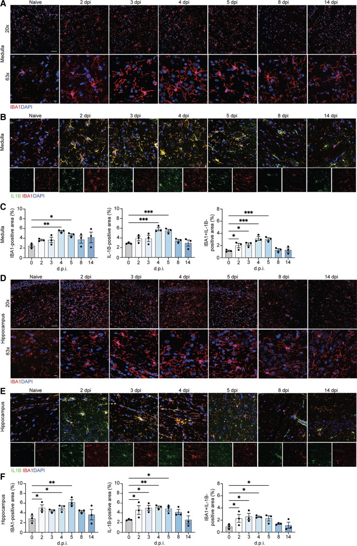
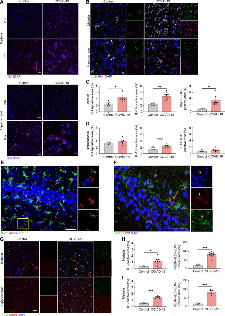
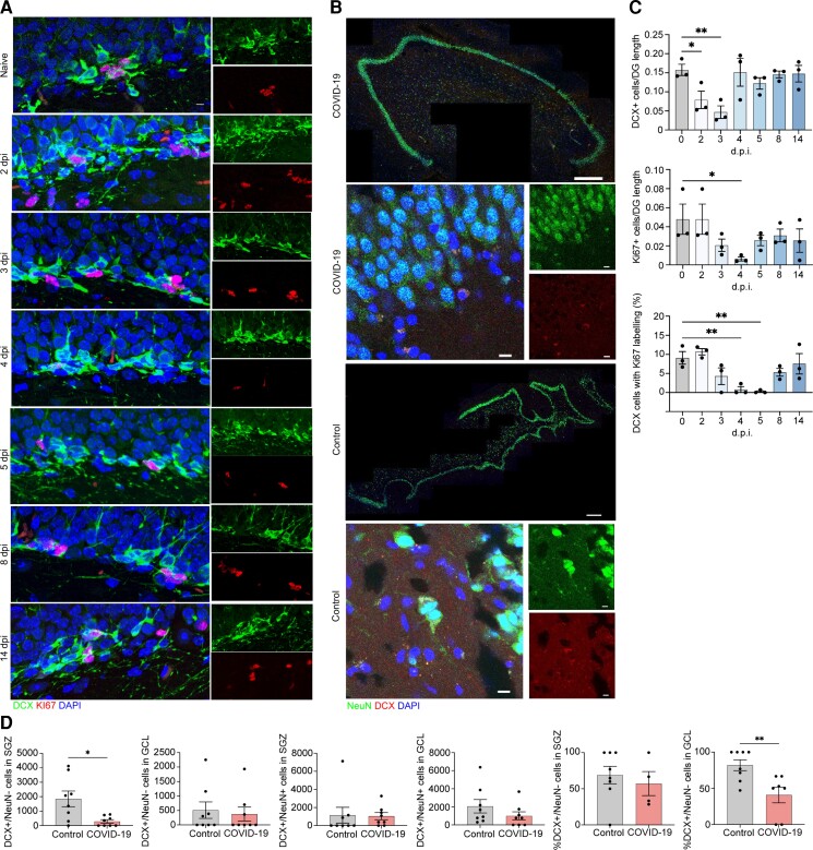
Infection with the severe acute respiratory syndrome coronavirus 2 (SARS-CoV-2) is associated with acute and postacute cognitive and neuropsychiatric symptoms including impaired memory, concentration, attention, sleep and affect. Mechanisms underlying these brain symptoms remain understudied. Here we report that SARS-CoV-2-infected hamsters exhibit a lack of viral neuroinvasion despite aberrant blood-brain barrier permeability. Hamsters and patients deceased from coronavirus disease 2019 (COVID-19) also exhibit microglial activation and expression of interleukin (IL)-1β and IL-6, especially within the hippocampus and the medulla oblongata, when compared with non-COVID control hamsters and humans who died from other infections, cardiovascular disease, uraemia or trauma. In the hippocampal dentate gyrus of both COVID-19 hamsters and humans, we observed fewer neuroblasts and immature neurons. Protracted inflammation, blood-brain barrier disruption and microglia activation may result in altered neurotransmission, neurogenesis and neuronal damage, explaining neuropsychiatric presentations of COVID-19. The involvement of the hippocampus may explain learning, memory and executive dysfunctions in COVID-19 patients.

Keywords: COVID-19; SARS-CoV-2; brain; cytokine; neurogenesis.


Figures

Figure 1
Figure 1
SARS-CoV2 infects hamster ONE and induces BBB disruption in hamsters and patients with COVID-19. (A) In situ hybridization for viral RNA at 2 dpi revealed SARS-CoV2 consistently targeted the ethmoturbinates of hamsters, with no infection of the CNS parenchyma. (B) Representative images of viral SARS-CoV2 mRNA in the hamster ethmoturbinates at naïve, 2, 3, 4, 5, 8 and 14 dpi. (C) Co-localization of viral RNA (red) via in situ hybridization and immunodetection of K18+ sustenticular cells (green) of the ONE in naïve or SARS-CoV2-infected hamsters at 7 dpi. Nuclei counterstained with DAPI (blue). (D) Representative image of blood–brain permeability in the hamster brain 2 dpi, showing staining for IgG (green) and DAPI (blue). (E) Representative images of IgG detection (green) within hamster MO and hippocampi at naïve, 2, 3, 4, 5, 8 and 14 dpi, and nuclear stain, DAPI (blue), followed by (F) quantitation of IgG intensity in the total CNS parenchyma (white outline) (top, Overview), medulla (middle, Medulla), and hippocampus (bottom, Hippocampus) at all time points. (G) Representative image of blood–brain permeability in the medulla (left) and hippocampus (right) of control and COVID-19 patient tissue, depicting detection of fibrinogen (green) and DAPI (blue). (H) Quantification of fibrinogen intensity in control versus COVID-19 patient tissues derived from medulla (top) and hippocampus (bottom). Data were pooled from at least two independent experiments. Scale bars = 50 μm (×10), 20 μm (×20) or 10 μm (×63). Data represent the mean ± SEM and were analysed by two-way ANOVA or Student’s t-test.
Figure 2
Figure 2
Microglia contribute to neuroinflammation in the medulla oblongata and hippocamus of SARS-CoV-2-infected hamsters. Representative images of IBA1 in the hamster inferior olivary nuclei (ION) (A) and hippocampus (D) at naïve, 2, 3, 4, 5, 8 and 14 dpi, showing staining for IBA1 (red) and DAPI (blue) at ×20 and ×63. Immunostaining for IL-1β and IBA1 in the hamster ION (B) and hippocampi (E) at naïve, 2, 3, 4, 5, 8 and 14 dpi, presented as microscopy with IBA1 (red), IL-1β (green) and DAPI (blue). Quantitation of per cent IBA1+ and IL-1β+ areas, and IL-1β+IBA1+ area, normalized to total IL-1B+ area for ION (C) and hamster (F). Data were pooled from at least two independent experiments. Scale bars = 20 μm (×20) or 10 μm (×63). Data represent the mean ± SEM and were analysed by two-way ANOVA or Student’s t-test.
Figure 3
Figure 3
Microglia and neurons contribute to neuroinflammation in the medulla and hippocampus of COVID-19 patients. Representative image of IBA1 in control and COVID-19 patient ION (A, top) and hippocampus (A, bottom), showing staining for IBA1 (magenta) and DAPI (blue) at ×20 and ×63, and quantified for per cent IBA1+ area (C and D, left). Immunostaining for IL-1β and IBA1 in ION and hippocampus of control and COVID-19 patients, presented as microscopy with IBA1 (magenta), IL-1β (green) and DAPI (blue) (B) and per cent IL-1β+ area and IL-1β+IBA1+ area, normalized to total IBA1+ areas for both regions (C and D, middle and right). (E) Representative images of IBA1 in the human adult hippocampus with high-magnification images single channel. Sections stained with DAPI (blue), IBA1 (green) and DCX (red) in non-COVID-19 control. Scale bar = 25 μm. (F) Representative images of GFAP in the human adult hippocampus with high-magnification images single channel. Sections stained with DAPI (blue), GFAP (green) and DCX (red) in non-COVID-19 control. The arrow points to a single DCX+/GFAP neuron in the subgranular zone (SGZ). Scale bar = 25 μm. Immunostaining for IL-6 and NeuN in ION and hippocampus of control and COVID-19 patients, presented as microscopy with NeuN (red), IL-6 (green) and DAPI (blue) (G) and per cent IL-6+ area and IL-16+NeuN+ area, normalized to total NeuN+ area (H and I). Data were pooled from at least two independent experiments. Scale bars = 20 μm (×20) or 10 μm (×63). Data represent the mean ± SEM and were analysed by two-way ANOVA or Student’s t-test.
Figure 4
Figure 4
Neuroblast proliferation in the SARS-CoV2 infected hamster and doublecortin (DCX)-positive cells and neurons in human hippocampus from COVID-19 patient and non-COVID-19 control. (A) Microscopy of the dentate gyrus of hamsters at naïve, 2, 3, 4, 5, 8 and 14 dpi, showing staining of Ki67 (red), neuroblast (green) and DAPI (blue), followed by quantification of per cent Ki67+DCX+ cells, normalized to the total number of DCX+ cells. (C) Quantification of per cent DCX+Ki67+ area, normalized to total DCX+ area in the rostral migratory stream of hamsters at naïve, 2, 3, 4, 5, 8 and 14 dpi. Data were pooled from at least two independent experiments. Scale bars = 50 μm. Data represent the mean ± SEM and were analysed by two-way ANOVA. (B) Select images of whole hippocampus and high-magnification images sections stained with DAPI (blue), NeuN (green) and DCX (red) from COVID-19 patient and non-COVID-19 control. The granule cell layer (GCL), subgranular zone (SGZ) and molecular layer (ML) of the dentate gyrus are visible, combined channels imaged at ×20; scale bar = 500 μm. High-magnification images were captured at ×63, scale bar = 20 μm. (D) In the SGZ, DCX+/NeuN cells were fewer in COVID-19 patients versus controls [P = 0.026; t(7.794) = 2.731; Welch’s t-test for non-stoichiometric data], with no group differences in DCX+/NeuN+ cell number (P = 0.189; Mann–Whitney). In the GCL, neither DCX+/NeuN cell count (P = 0.846; Mann–Whitney) nor DCX+/NeuN+ cell count (P = 0.378; Mann–Whitney) differs between COVID-19 and control subjects. Per cent of DCX+/NeuN cells located in the SGZ versus the GCL did not differ between control and COVID-19 groups (P = 0.453; Mann–Whitney). Per cent of DCX+/NeuN+ cells located in the GCL versus the SGZ was lower in COVID-19 patients versus control subjects (P = 0.009; Mann–Whitney).

Update of

References

    1. Jaywant A, Vanderlind WM, Alexopoulos GS, Fridman CB, Perlis RH, Gunning FM. Frequency and profile of objective cognitive deficits in hospitalized patients recovering from COVID-19. Neuropsychopharmacology. 2021;46:2235–2240. - PMC - PubMed
    1. Nalbandian A, Sehgal K, Gupta A, et al. Post-acute COVID-19 syndrome. Nat Med. 2021;27:601–615. - PMC - PubMed
    1. Helms J, Kremer S, Merdji H, et al. Neurologic features in severe SARS-CoV-2 infection. N Engl J Med. 2020;382:2268–2270. - PMC - PubMed
    1. Mendez R, Balanza-Martinez V, Luperdi SC, et al. Long-term neuropsychiatric outcomes in COVID-19 survivors: A 1-year longitudinal study. J Intern Med. 2022;291:247–251. - PMC - PubMed
    1. Thakur KT, Miller EH, Glendinning MD, et al. COVID-19 neuropathology at Columbia University Irving Medical Center/New York Presbyterian Hospital. Brain. 2021;144:2696–2708. - PMC - PubMed

Publication types