

Development of a New Radiation Shield for the Face and Neck of IVR Physicians
- PMID: 36004878
- PMCID: PMC9404996
- DOI: 10.3390/bioengineering9080354
Development of a New Radiation Shield for the Face and Neck of IVR Physicians
Abstract
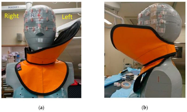
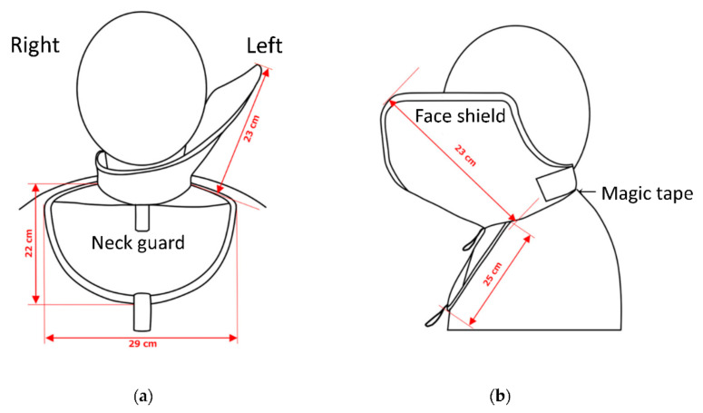
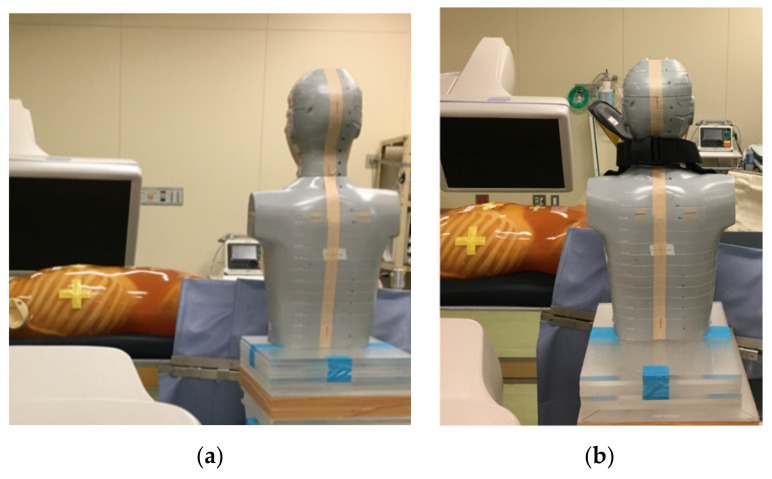
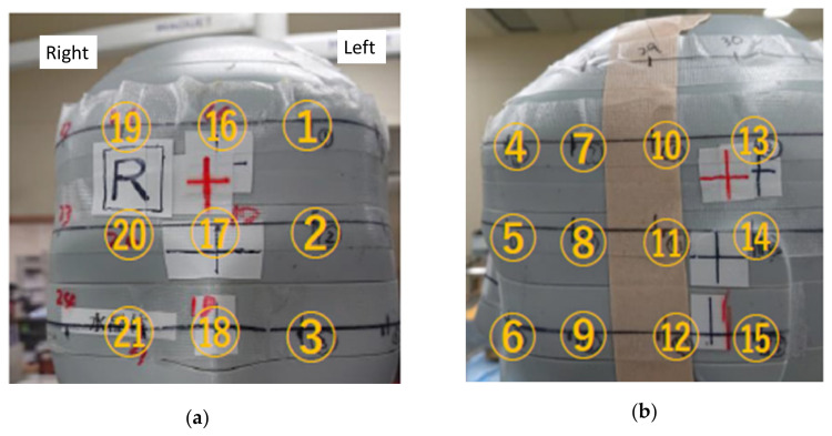
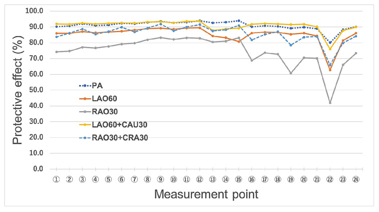
Interventional radiology (IVR) procedures are associated with increased radiation exposure and injury risk. Furthermore, radiation eye injury (i.e., cataract) in IVR staff have also been reported. It is crucial to protect the eyes of IVR physicians from X-ray radiation exposure. Many IVR physicians use protective Pb eyeglasses to reduce occupational eye exposure. However, the shielding effects of Pb eyeglasses are inadequate. We developed a novel shield for the face (including eyes) of IVR physicians. The novel shield consists of a neck and face guard (0.25 mm Pb-equivalent rubber sheet, nonlead protective sheet). The face shield is positioned on the left side of the IVR physician. We assessed the shielding effects of the novel shield using a phantom in the IVR X-ray system; a radiophotoluminescence dosimeter was used to measure the radiation exposure. In this phantom study, the effectiveness of the novel device for protecting against radiation was greater than 80% in almost all measurement situations, including in terms of eye lens exposure. A large amount of scattered radiation reaches the left side of IVR physicians. The novel radiation shield effectively protects the left side of the physician from this scattered radiation. Thus, the device can be used to protect the face and eyes of IVR physicians from occupational radiation exposure. The novel device will be useful for protecting the face (including eyes) of IVR physicians from radiation, and thus could reduce the rate of radiation injury. Based on the positive results of this phantom study, we plan to perform a clinical experiment to further test the utility of this novel radiation shield for IVR physicians.
Keywords: X-ray examination; disaster medicine; face shield; fluoroscopically guided interventional procedures; fluoroscopy; interventional radiology (IVR); percutaneous coronary intervention (PCI); protective apron; radiation dose; radiation protection and safety.
Conflict of interest statement
The authors declare no conflict of interest.
Figures
References
-
- Haga Y., Chida K., Sota M., Kaga Y., Abe M., Inaba Y., Suzuki M., Meguro T., Zuguchi M. Hybrid operating room system for the treatment of thoracic and abdominal aortic aneurysms: Evaluation of the radiation dose received by patients. Diagnostics. 2020;10:846. doi: 10.3390/diagnostics10100846. - DOI - PMC - PubMed
-
- Chida K., Saito H., Otani H., Kohzuki M., Takahashi S., Yamada S., Shirato K., Zuguchi M. Relationship between fluoroscopic time, dose—Area product, body weight, and maximum radiation skin dose in cardiac interventional procedures. Am. J. Roentgenol. 2006;186:774–778. doi: 10.2214/AJR.04.1653. - DOI - PubMed
Grants and funding
LinkOut - more resources
Full Text Sources
Miscellaneous
