. 2022 Aug 4;12(8):596.
doi: 10.3390/bios12080596.

Probing the Hepatitis B Virus E-Antigen with a Nanopore Sensor Based on Collisional Events Analysis

Affiliations

Probing the Hepatitis B Virus E-Antigen with a Nanopore Sensor Based on Collisional Events Analysis

Ioana C Bucataru et al. Biosensors (Basel). .

Abstract

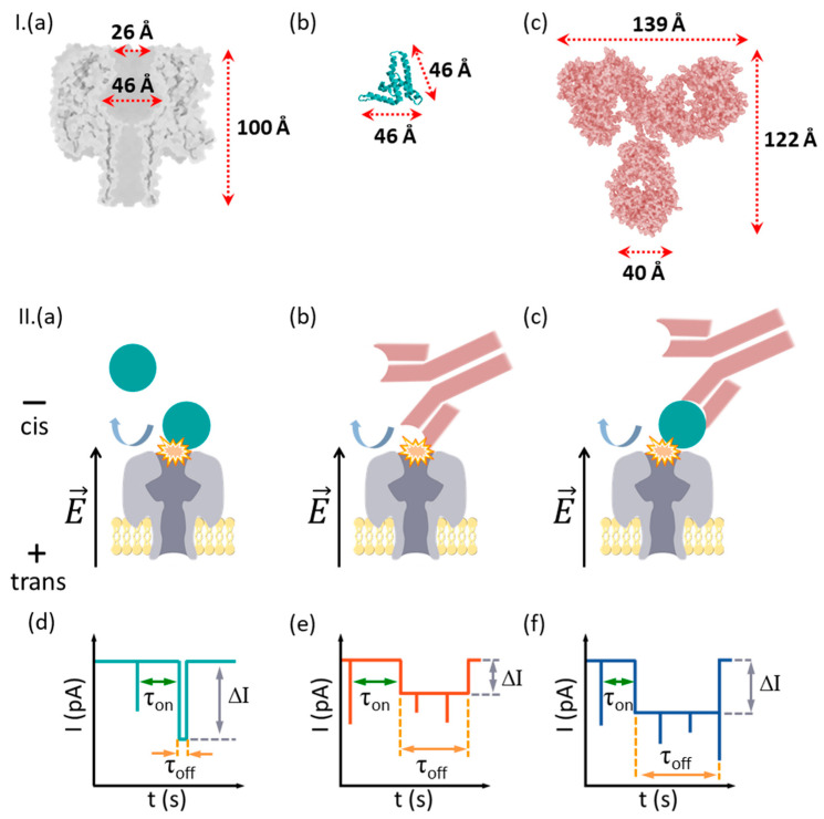
Real-time monitoring, simple operation, and cheaper methods for detecting immunological proteins hold the potential for a solid influence on proteomics and human biology, as they can promote the onset of timely diagnoses and adequate treatment protocols. In this work we present an exploratory study suggesting the applicability of resistive-pulse sensing technology in conjunction with the α-hemolysin (α-HL) protein nanopore, for the detection of the chronic hepatitis B virus (HBV) e-antigen (HBeAg). In this approach, the recognition between HBeAg and a purified monoclonal hepatitis B e antibody (Ab(HBeAg)) was detected via transient ionic current spikes generated by partial occlusions of the α-HL nanopore by protein aggregates electrophoretically driven toward the nanopore's vestibule entrance. Despite the steric hindrance precluding antigen, antibody, or antigen-antibody complex capture inside the nanopore, their stochastic bumping with the nanopore generated clear transient blockade events. The subsequent analysis suggested the detection of protein subpopulations in solution, rendering the approach a potentially valuable label-free platform for the sensitive, submicromolar-scale screening of HBeAg targets.

Keywords: antigen; electrophysiology; hepatitis B; monoclonal antibody; nanopore; single molecule detection.


Conflict of interest statement

The authors declare no conflict of interest.

Figures

Figure 1
Figure 1
Simplified description of the measurement principle. (I) In (ac) are presented at a similar scale the homo-heptameric α-HL protein nanopore, HBeAg protein antigen and its monoclonal antibody Ab(HBeAg). (II) Due to the exclusion volume effects, the nanopore cannot capture and accommodate inside it the HBeAg (a), Ab(HBeAg) antibody (b), or HBeAg–Ab(HBeAg) complexes formed in solution (c). The electrophoresis of anionic HBeAg, Ab(HBeAg) antibody, or HBeAg–Ab(HBeAg) complexes toward the nanopore result in collisional interactions with the nanopore, seen as stochastic spikes in the ionic current (df), correlated with the transient obturation of the α-HL’s vestibule entrance area by an incoming analyte. Such events are characterized by the blockade depth (ΔI), dwell time (τoff), and inter-events intervals (τon). In certain cases, a series of fast-occurring spikes are seen during a single collisional event (see schematics in panels e and f, the capture events), which we posit to reflect stochastic, spatial re-arrangements of the captured analyte at the nanopore’s mouth.
Figure 2
Figure 2
Despite their relative large size, the interactions between the cis-side-added HBeAg and Ab(HBeAg) with the α-HL nanopore are visible in electrophysiology experiments. (I) Reversible collisional interactions between the positively biased α-HL (a) and the cis-side-added HBeAg, present at 50 nM (b), 100 nM (c), and 150 nM (d) are seen as downwardly oriented spikes. The expanded traces illustrate the degree of heterogeneity of the blockade events, suggestive of geometrical re-orientations or/and structural changes of the captured analyte during individual bumping events. (II) The ionic current through a positively biased nanopore (a) is transiently blocked when the monoclonal antibody Ab(HBeAg) is added on the cis side of the chamber at incremental concentrations of 50 nM (b), 100 nM (c), and 200 nM (d), as a result of transient occlusions of the vestibule entrance of the nanopore during collisions. As presented in the expanded traces, the rich diversity of blockade substates manifested during individual collisional events, all characterized by various τoff values, supports the hypothesis of a highly dynamic state of the transiently captured antibody in terms of orientation and/or tertiary structure, all leading to various degrees of nanopore occlusion. The traces were recorded at an applied potential ΔV = +180mV in a 2 M KCl electrolyte solution, buffered at pH = 8 with a 10 mM HEPES solution.
Figure 3
Figure 3
Quantitative analysis of HbeAg–α-HL reversible interactions. (a) Concentration dependence of the time intervals reflecting the purified HbeAg-α-HL association (τon; a) and, respectively, dissociation (τoff; b), measured at an applied potential ΔV = +180 mV.
Figure 4
Figure 4
Detection of HBeAg–Ab(HBeAg) complexes with the α-HL nanopore. (ac) Interaction between α-HL and nonincubated, cis-side-added HBeAg and Ab(HBeAg) mixed at a 1:1 (100 nM:100 nM) and 1:2 (100 nM:200 nM) molar ratio generate solid ionic currents blockades across the open α-HL nanopore at an applied voltage ΔV = + 180 mV. (d) All-points histograms showing the distinct conductive states of α-HL nanopore, while interacting with the cis-side-added HbeAg, Ab(HBeAg), or HBeAg–Ab(HBeAg) complexes (1:1 molar ratio). In the inset we represent a zoomed-in excerpt evidencing the experimental shift in the open pore current through the nanopore in either case (see also text). (e) Shift-corrected histograms, all aligned to a similar value corresponding to the open nanopore current. (f) The shift-corrected, all-points histograms transformed in terms of percent relative current blockades (% IIo, where ∆I is the current magnitude of a blockade substate relative to the open nanopore current Io). (g) All-points histograms showing the distinct conductive states of α-HL nanopore, while interacting with the cis-side-added HbeAg, Ab(HBeAg), or HbeAg–Ab(HBeAg) complexes (1:2 molar ratio). As above, in the inset we represent an excerpt displaying the experimental variability in the open pore current through the nanopore during such measurements. (h) Shift-corrected histograms, all aligned to a similar value corresponding to the open nanopore current. (i) The shift-corrected, all-points histograms transformed in terms of percent relative current blockades (% IIo).
Figure 5
Figure 5
The noise analysis of collisional events ensued by proteins interacting with the α-HL nanopore (a) Representative power-spectra of the ionic current fluctuations recorded at ΔV = +180 mV, associated with the HbeAg, Ab(HBeAg), or HbeAg–Ab(HBeAg) complex collisions with a single α-HL nanopore. (b) The power spectrum of current fluctuations induced by HbeAg–Ab(HBeAg) complexes interacting with the α-HL nanopore remained unaltered in the presence of 1% human serum (HS). The yellow domains represent the frequency bandwidth containing excessive low-frequency noise contributions, subsequently excluded from the analysis (see also main text).
Figure 6
Figure 6
HbeAg–Ab(HBeAg) complex detections with a single α-HL nanopore in the presence of human serum proteins. Representative, original traces and related all-points histograms of the ionic current measured at ΔV = +180 mV across an open nanopore (a,b), then in the presence of cis-added Ag(HBe) antigen (100 nM) (c,d), followed by addition of the Ab(HBe) antibody (200 nM) (e,f), and subsequent pipetting of human serum (HS) at a 0.5% (v/v) (g,h) and, respectively, 1% (v/v) concentration (i,j). In certain cases, the HS addition led to the non-specific occlusion of the nanopore (red rectangle in panels (g,i), followed in certain cases by lipid membrane rupture.

Similar articles

Cited by

References

    1. Kasianowicz J.J., Brandin E., Branton D., Deamer D.W. Characterization of Individual Polynucleotide Molecules Using a Membrane Channel. Proc. Natl. Acad. Sci. USA. 1996;93:13770–13773. doi: 10.1073/pnas.93.24.13770. - DOI - PMC - PubMed
    1. Branton D., Deamer D.W., Marziali A., Bayley H., Benner S.A., Butler T., Di Ventra M., Garaj S., Hibbs A., Huang X., et al. The Potential and Challenges of Nanopore Sequencing. Nat. Biotechnol. 2008;26:1146–1153. doi: 10.1038/nbt.1495. - DOI - PMC - PubMed
    1. Cao C., Ying Y.-L., Hu Z.-L., Liao D.-F., Tian H., Long Y.-T. Discrimination of Oligonucleotides of Different Lengths with a Wild-Type Aerolysin Nanopore. Nat. Nanotechnol. 2016;11:713–718. doi: 10.1038/nnano.2016.66. - DOI - PubMed
    1. Stoddart D., Heron A.J., Mikhailova E., Maglia G., Bayley H. Single-Nucleotide Discrimination in Immobilized DNA Oligonucleotides with a Biological Nanopore. Proc. Natl. Acad. Sci. USA. 2009;106:7702–7707. doi: 10.1073/pnas.0901054106. - DOI - PMC - PubMed
    1. Howorka S., Cheley S., Bayley H. Sequence-Specific Detection of Individual DNA Strands Using Engineered Nanopores. Nat. Biotechnol. 2001;19:636–639. doi: 10.1038/90236. - DOI - PubMed

LinkOut - more resources