

Cardiac and Respiratory Influences on Intracranial and Neck Venous Flow, Estimated Using Real-Time Phase-Contrast MRI
- PMID: 36005008
- PMCID: PMC9405895
- DOI: 10.3390/bios12080612
Cardiac and Respiratory Influences on Intracranial and Neck Venous Flow, Estimated Using Real-Time Phase-Contrast MRI
Abstract
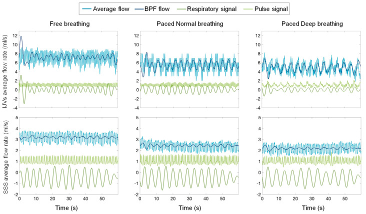
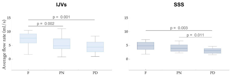
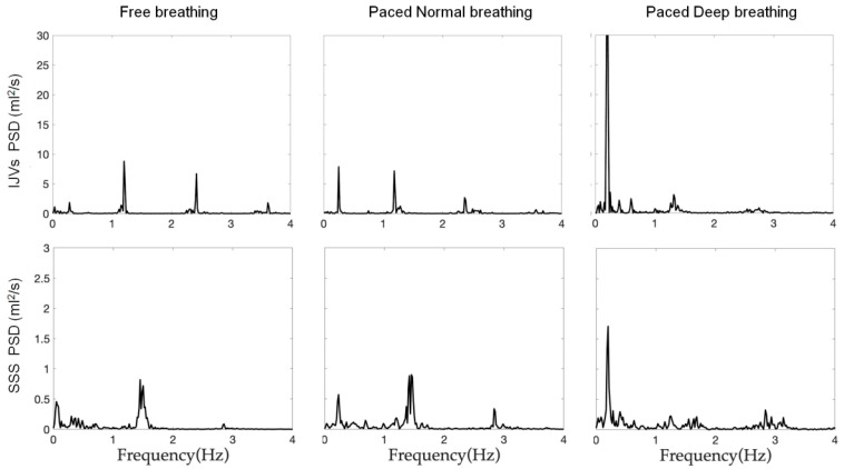
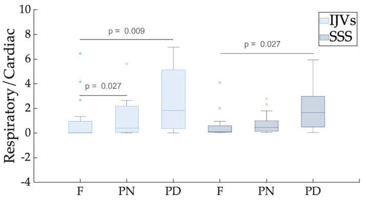
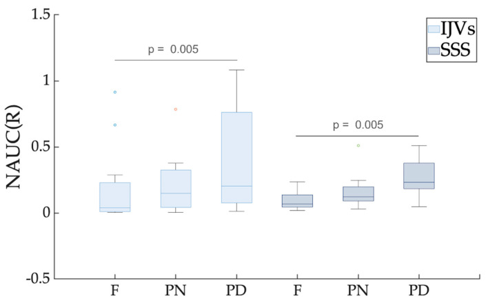
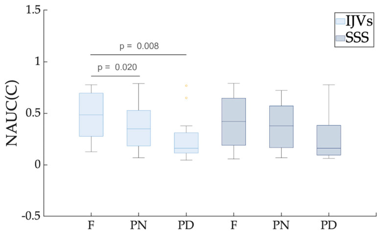
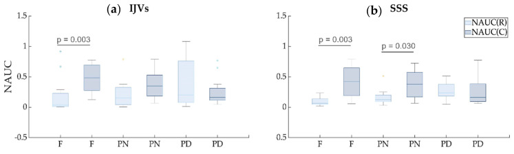
The study of brain venous drainage has gained attention due to its hypothesized link with various neurological conditions. Intracranial and neck venous flow rate may be estimated using cardiac-gated cine phase-contrast (PC)-MRI. Although previous studies showed that breathing influences the neck's venous flow, this aspect could not be studied using the conventional segmented PC-MRI since it reconstructs a single cardiac cycle. The advent of real-time PC-MRI has overcome these limitations. Using this technique, we measured the internal jugular veins and superior sagittal sinus flow rates in a group of 16 healthy subjects (12 females, median age of 23 years). Comparing forced-breathing and free-breathing, the average flow rate decreased and the respiratory modulation increased. The flow rate decrement may be due to a vasoreactive response to deep breathing. The respiratory modulation increment is due to the thoracic pump's greater effect during forced breathing compared to free breathing. These results showed that the breathing mode influences the average blood flow and its pulsations. Since effective drainage is fundamental for brain health, rehabilitative studies might use the current setup to investigate if respiratory exercises positively affect clinical variables and venous drainage.
Keywords: RT-PC MRI; intracranial venous flow; neck venous flow.
Conflict of interest statement
The authors declare no conflict of interest. The funders had no role in the design of the study; in the collection, analyses, or interpretation of data; in the writing of the manuscript, or in the decision to publish the results.
Figures
Similar articles
-
The Effects of Free Breathing on Cerebral Venous Flow: A Real-Time Phase Contrast MRI Study in Healthy Adults.J Neurosci. 2024 Jan 17;44(3):e0965232023. doi: 10.1523/JNEUROSCI.0965-23.2023. J Neurosci. 2024. PMID: 37968115 Free PMC article.
-
Blood and cerebrospinal fluid flow oscillations measured with real-time phase-contrast MRI: breathing mode matters.Fluids Barriers CNS. 2022 Dec 14;19(1):100. doi: 10.1186/s12987-022-00394-0. Fluids Barriers CNS. 2022. PMID: 36517859 Free PMC article.
-
Validating the accuracy of real-time phase-contrast MRI and quantifying the effects of free breathing on cerebrospinal fluid dynamics.Fluids Barriers CNS. 2024 Mar 7;21(1):25. doi: 10.1186/s12987-024-00520-0. Fluids Barriers CNS. 2024. PMID: 38454518 Free PMC article.
-
Quantifying the influence of respiration and cardiac pulsations on cerebrospinal fluid dynamics using real-time phase-contrast MRI.J Magn Reson Imaging. 2017 Aug;46(2):431-439. doi: 10.1002/jmri.25591. Epub 2017 Feb 2. J Magn Reson Imaging. 2017. PMID: 28152239
-
Brain blood and cerebrospinal fluid flow dynamics during rhythmic handgrip exercise in young healthy men and women.J Physiol. 2021 Mar;599(6):1799-1813. doi: 10.1113/JP281063. Epub 2021 Feb 16. J Physiol. 2021. PMID: 33481257
Cited by
-
In Vivo Biomarker Imaging: Paving the Way for Precision Medicine.Biosensors (Basel). 2023 Apr 3;13(4):454. doi: 10.3390/bios13040454. Biosensors (Basel). 2023. PMID: 37185529 Free PMC article.
-
Neurofluids and the glymphatic system: anatomy, physiology, and imaging.Br J Radiol. 2023 Nov;96(1151):20230016. doi: 10.1259/bjr.20230016. Epub 2023 Jun 1. Br J Radiol. 2023. PMID: 37191063 Free PMC article. Review.
-
Quantification of the Dynamics of the Vascular Flows in the Cerebral Arterial and Venous Trees.Biomedicines. 2025 May 1;13(5):1106. doi: 10.3390/biomedicines13051106. Biomedicines. 2025. PMID: 40426933 Free PMC article.
-
The Effects of Free Breathing on Cerebral Venous Flow: A Real-Time Phase Contrast MRI Study in Healthy Adults.J Neurosci. 2024 Jan 17;44(3):e0965232023. doi: 10.1523/JNEUROSCI.0965-23.2023. J Neurosci. 2024. PMID: 37968115 Free PMC article.
-
Respiratory influence on cerebral blood flow and blood volume - A 4D flow MRI study.J Cereb Blood Flow Metab. 2025 Jan 30:271678X251316395. doi: 10.1177/0271678X251316395. Online ahead of print. J Cereb Blood Flow Metab. 2025. PMID: 39883800 Free PMC article.
References
MeSH terms
Grants and funding
LinkOut - more resources
Full Text Sources
Medical
