

Electrochemical DNA Biosensor Based on Mercaptopropionic Acid-Capped ZnS Quantum Dots for Determination of the Gender of Arowana Fish
- PMID: 36005045
- PMCID: PMC9405751
- DOI: 10.3390/bios12080650
Electrochemical DNA Biosensor Based on Mercaptopropionic Acid-Capped ZnS Quantum Dots for Determination of the Gender of Arowana Fish
Abstract
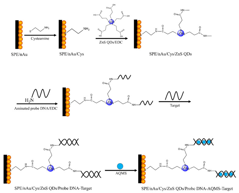
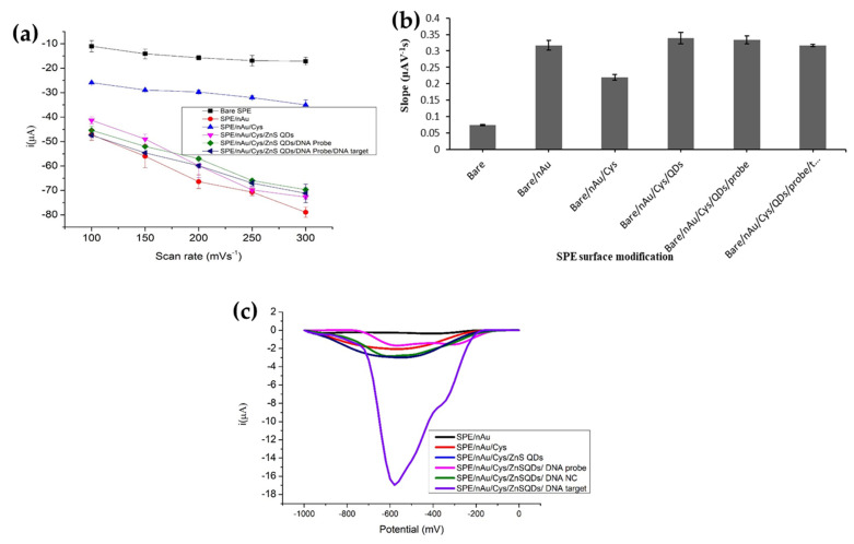
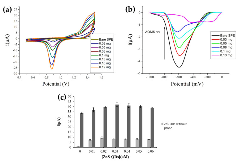
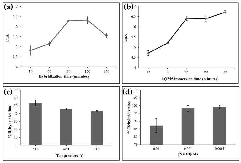
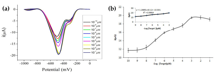
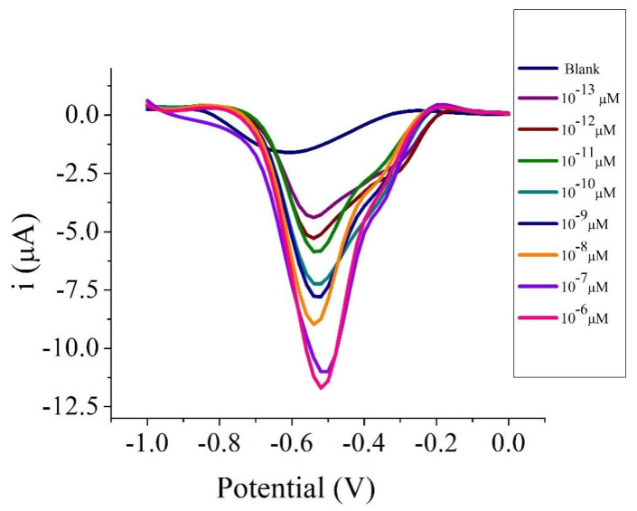
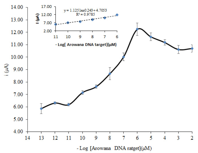
A new electrochemical DNA biosensor based on mercaptopropionic acid (MPA)-capped ZnS quantum dots (MPA-ZnS QDs) immobilization matrix for covalent binding with 20-base aminated oligonucleotide has been successfully developed. Prior to the modification, screen-printed carbon paste electrode (SPE) was self-assembled with multilayer gold nanoparticles (AuNPs) and cysteamine (Cys). The inclusion of MPA-ZnS QDs semiconducting material in modified electrodes has enhanced the electron transfer between the SPE transducer and DNA leading to improved bioanalytical assay of target biomolecules. Electrochemical studies performed by cyclic voltammetry (CV) and differential pulsed voltammetry (DPV) demonstrated that the MPA-ZnS QDs modified AuNPs electrode was able to produce a lower charge transfer resistance response and hence higher electrical current response. Under optimal conditions, the immobilized synthetic DNA probe exhibited high selectivity towards synthetic target DNA. Based on the DPV response of the reduction of anthraquinone monosulphonic acid (AQMS) redox probe, the MPA-ZnS QDs-based electrochemical DNA biosensor responded to target DNA concentration from 1 × 10⁻⁹ μM to 1 × 10⁻³ μM with a sensitivity 1.2884 ± 0.12 µA, linear correlation coefficient (R2) of 0.9848 and limit of detection (LOD) of 1 × 10-11 μM target DNA. The DNA biosensor exhibited satisfactory reproducibility with an average relative standard deviation (RSD) of 7.4%. The proposed electrochemical transducer substrate has been employed to immobilize the aminated Arowana fish (Scleropages formosus) DNA probe. The DNA biosensor showed linearity to target DNA from 1 × 10-11 to 1 × 10⁻⁶ µM (R2 = 0.9785) with sensitivity 1.1251 ± 0.243 µA and LOD of 1 × 10-11 µM. The biosensor has been successfully used to determine the gender of Arowana fish without incorporating toxic raw materials previously employed in the hazardous processing conditions of polypyrrole chemical conducting polymer, whereby the cleaning step becomes difficult with thicker films due to high levels of toxic residues from the decrease in polymerization efficacy as films grew.
Keywords: DNA biosensor; ZnS QDs; electrochemistry; gold nanoparticles; screen printed electrode.
Conflict of interest statement
The authors declare no conflict of interest.
Figures
Similar articles
-
A Highly Sensitive Electrochemical DNA Biosensor from Acrylic-Gold Nano-composite for the Determination of Arowana Fish Gender.Nanoscale Res Lett. 2017 Aug 10;12(1):484. doi: 10.1186/s11671-017-2254-y. Nanoscale Res Lett. 2017. PMID: 28798991 Free PMC article.
-
Blue-emitting SiO2-coated Si-doped ZnSeS quantum dots conjugated aptamer-molecular beacon as an electrochemical and metal-enhanced fluorescence biosensor for SARS-CoV-2 spike protein.Anal Chim Acta. 2023 Nov 15;1281:341926. doi: 10.1016/j.aca.2023.341926. Epub 2023 Oct 17. Anal Chim Acta. 2023. PMID: 39492217
-
Nanocrystalline cellulose decorated quantum dots based tyrosinase biosensor for phenol determination.Mater Sci Eng C Mater Biol Appl. 2019 Jun;99:37-46. doi: 10.1016/j.msec.2019.01.082. Epub 2019 Jan 21. Mater Sci Eng C Mater Biol Appl. 2019. PMID: 30889711
-
Beyond traditional biosensors: Recent advances in gold nanoparticles modified electrodes for biosensing applications.Talanta. 2024 Feb 1;268(Pt 1):125280. doi: 10.1016/j.talanta.2023.125280. Epub 2023 Oct 12. Talanta. 2024. PMID: 37862755 Review.
-
Substrate Materials for Biomolecular Immobilization within Electrochemical Biosensors.Biosensors (Basel). 2021 Jul 15;11(7):239. doi: 10.3390/bios11070239. Biosensors (Basel). 2021. PMID: 34356710 Free PMC article. Review.
Cited by
-
An Ultrasensitive Voltammetric Genosensor for the Detection of Bacteria Vibrio cholerae in Vegetable and Environmental Water Samples.Biosensors (Basel). 2023 Jun 4;13(6):616. doi: 10.3390/bios13060616. Biosensors (Basel). 2023. PMID: 37366981 Free PMC article.
-
Recent Developments in the Design and Fabrication of Electrochemical Biosensors Using Functional Materials and Molecules.Biosensors (Basel). 2023 Mar 27;13(4):424. doi: 10.3390/bios13040424. Biosensors (Basel). 2023. PMID: 37185499 Free PMC article. Review.
-
Strategies in the optimization of DNA hybridization conditions and its role in electrochemical detection of dengue virus (DENV) using response surface methodology (RSM).RSC Adv. 2023 Jun 16;13(27):18748-18759. doi: 10.1039/d3ra00216k. eCollection 2023 Jun 15. RSC Adv. 2023. PMID: 37362605 Free PMC article.
References
-
- Cai J., Sun B., Gou X., Gou Y., Li W., Hu F. A novel way for analysis of calycosin via polyaniline functionalized graphene quantum dots fabricated electrochemical sensor. J. Electroanal. Chem. 2018;816:123–131. doi: 10.1016/j.jelechem.2018.03.035. - DOI
-
- Esmaeili C., Heng L.Y., Chiang C.P., Rashid Z.A., Safitri E., Malon Marugan R.S.P. A DNA biosensor based on kappa-carrageenan-polypyrrole-gold nanoparticles composite for gender determination of Arowana fish (Scleropages formosus) Sens. Actuators B Chem. 2017;242:616–624. doi: 10.1016/j.snb.2016.11.061. - DOI
MeSH terms
Substances
LinkOut - more resources
Full Text Sources
