

Multi-Factors Cooperatively Actuated Photonic Hydrogel Aptasensors for Facile, Label-Free and Colorimetric Detection of Lysozyme
- PMID: 36005058
- PMCID: PMC9406194
- DOI: 10.3390/bios12080662
Multi-Factors Cooperatively Actuated Photonic Hydrogel Aptasensors for Facile, Label-Free and Colorimetric Detection of Lysozyme
Abstract
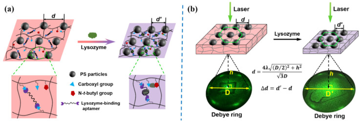
Responsive two-dimensional photonic crystal (2DPC) hydrogels have been widely used as smart sensing materials for constructing various optical sensors to accurately detect different target analytes. Herein, we report photonic hydrogel aptasensors based on aptamer-functionalized 2DPC poly(acrylamide-acrylic acid-N-tert-butyl acrylamide) hydrogels for facile, label-free and colorimetric detection of lysozyme in human serum. The constructed photonic hydrogel aptasensors undergo shrinkage upon exposure to lysozyme solution through multi-factors cooperative actuation. Here, the specific binding between the aptamer and lysozyme, and the simultaneous interactions between carboxyl anions and N-tert-butyl groups with lysozyme, increase the cross-linking density of the hydrogel, leading to its shrinkage. The aptasensors' shrinkage decreases the particle spacing of the 2DPC embedded in the hydrogel network. It can be simply monitored by measuring the Debye diffraction ring of the photonic hydrogel aptasensors using a laser pointer and a ruler without needing sophisticated apparatus. The significant shrinkage of the aptasensors can be observed by the naked eye via the hydrogel size and color change. The aptasensors show good sensitivity with a limit of detection of 1.8 nM, high selectivity and anti-interference for the detection of lysozyme. The photonic hydrogel aptasensors have been successfully used to accurately determine the concentration of lysozyme in human serum. Therefore, novel photonic hydrogel aptasensors can be constructed by designing functional monomers and aptamers that can specifically bind target analytes.
Keywords: aptamer conformational change; colorimetric sensing; multi-factors actuation; photonic hydrogel aptasensors; volume phase transition.
Conflict of interest statement
The authors declare no conflict of interest.
Figures


Similar articles
-
Aptamer-functionalized 2D photonic crystal hydrogels for detection of adenosine.Mikrochim Acta. 2022 Oct 15;189(11):418. doi: 10.1007/s00604-022-05521-0. Mikrochim Acta. 2022. PMID: 36242658
-
DNA-Crosslinked 2D Photonic Crystal Hydrogels for Detection of Adenosine Actuated by an Adenosine-Binding Aptamer.ACS Sens. 2022 Jun 24;7(6):1648-1656. doi: 10.1021/acssensors.1c02424. Epub 2022 May 27. ACS Sens. 2022. PMID: 35623053
-
Responsive photonic hydrogel for colorimetric detection of formaldehyde.Spectrochim Acta A Mol Biomol Spectrosc. 2023 Nov 5;300:122920. doi: 10.1016/j.saa.2023.122920. Epub 2023 May 30. Spectrochim Acta A Mol Biomol Spectrosc. 2023. PMID: 37269656
-
Advances in photonic crystal hydrogels for biomedical research: A review.J Biotechnol. 2025 Aug;404:162-174. doi: 10.1016/j.jbiotec.2025.04.019. Epub 2025 Apr 28. J Biotechnol. 2025. PMID: 40306327 Review.
-
Aptasensors for lysozyme detection: Recent advances.Talanta. 2021 May 1;226:122169. doi: 10.1016/j.talanta.2021.122169. Epub 2021 Jan 30. Talanta. 2021. PMID: 33676711 Review.
Cited by
-
Stabilization mechanism and remediation effectiveness of Pb and cd in agricultural soil using nonmetallic minerals.Sci Rep. 2025 Apr 14;15(1):12757. doi: 10.1038/s41598-025-96970-z. Sci Rep. 2025. PMID: 40223021 Free PMC article.
-
Review on Carbon Dot-Based Fluorescent Detection of Biothiols.Biosensors (Basel). 2023 Mar 2;13(3):335. doi: 10.3390/bios13030335. Biosensors (Basel). 2023. PMID: 36979547 Free PMC article. Review.
-
A flexible magnetic DNA biosensor composed of AgNWs/hydrogel/PS/Fe3O4 for the detection of ASFV P72 protein gene fragment.Mikrochim Acta. 2025 Mar 28;192(4):268. doi: 10.1007/s00604-025-07114-z. Mikrochim Acta. 2025. PMID: 40153065
References
-
- Shen P., Zhang Y., Cai Z., Liu R., Xu X., Li R., Wang J.-J., Yang D. Three-Dimensional/Two-Dimensional Photonic Crystal Hydrogels for Biosensing. J. Mater. Chem. C. 2021;9:5840–5857. doi: 10.1039/D1TC00830G. - DOI
-
- Jang K., Horne W.S., Asher S.A. Human Serum Phenylpyruvate Quantification Using Responsive 2D Photonic Crystal Hydrogels via Chemoselective Oxime Ligation: Progress toward Developing Phenylalanine-Sensing Elements. ACS Appl. Mater. Interfaces. 2020;12:39612–39619. doi: 10.1021/acsami.0c08787. - DOI - PubMed
-
- Xue F., Meng Z., Wang F., Wang Q., Xue M., Xu Z. A 2-D Photonic Crystal Hydrogel for Selective Sensing of Glucose. J. Mater. Chem. A. 2014;2:9559–9565. doi: 10.1039/C4TA01031K. - DOI
MeSH terms
Substances
Grants and funding
LinkOut - more resources
Full Text Sources
