. 2022 Jul 29;8(8):477.
doi: 10.3390/gels8080477.

Dynamic and Self-Healable Chitosan/Hyaluronic Acid-Based In Situ-Forming Hydrogels

Affiliations

Dynamic and Self-Healable Chitosan/Hyaluronic Acid-Based In Situ-Forming Hydrogels

Sheila Maiz-Fernández et al. Gels. .

Abstract

In situ-forming, biodegradable, and self-healing hydrogels, which maintain their integrity after damage, owing to dynamic interactions, are essential biomaterials for bioapplications, such as tissue engineering and drug delivery. This work aims to develop in situ, biodegradable and self-healable hydrogels based on dynamic covalent bonds between N-succinyl chitosan (S-CHI) and oxidized aldehyde hyaluronic acid (A-HA). A robust effect of the molar ratio of both S-CHI and A-HA was observed on the swelling, mechanical stability, rheological properties and biodegradation kinetics of these hydrogels, being the stoichiometric ratio that which leads to the lowest swelling factor (×12), highest compression modulus (1.1·10−3 MPa), and slowest degradation (9 days). Besides, a rapid (3 s) self-repairing ability was demonstrated in the macro scale as well as by rheology and mechanical tests. Finally, the potential of these biomaterials was evidenced by cytotoxicity essay (>85%).

Keywords: N-succinyl chitosan; aldehyde hyaluronic acid; dynamic bonds; hydrogels; self-healing.


Conflict of interest statement

The authors declare no conflict of interest.

Figures

Figure 1
Figure 1
(A) Chemical structures of chitosan and N-succinyl-chitosan and their (B) FTIR and (C) 1H-NMR spectra.
Figure 2
Figure 2
(A) Chemical structures of hyaluronic acid and oxidized hyaluronic acid dialdehyde and their (B) FTIR and (C) 1H-NMR spectra.
Figure 3
Figure 3
(A) Gelation mechanism between N-succinyl chitosan and aldehyde hyaluronic acid by the Schiff’s base reaction. (B) FTIR spectra of the modified polysaccharides and the corresponding 5:5 S-CHI/A:HA hydrogel.
Figure 4
Figure 4
Swelling factors of the different hydrogels.
Figure 5
Figure 5
(A) Compressive tests, (B) Rheological frequency sweep measurements (filled circles G′ and open circles G″) and (C) Degradation kinetics of the S-CHI/A-HA hydrogels with different polysaccharide content.
Figure 6
Figure 6
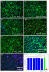
Biocompatibility of the hydrogels. Images of the cells after 24 h of contact with the hydrogels (AG) show good biocompatibility in all tested formulations. Quantification of the ratio between dead cells with red stained nuclei and all cells (with blue nuclei), showing no statistical differences between all conditions tested (H). (n.s. not significant). The positive control corresponds to cell membranes incubated in ice-cold ethanol being permeable to ethidium homodimer. Cells in the negative control were cultured without hydrogel.
Figure 7
Figure 7
(A) Self-healing of 1 cut-recovery cycle. Compression strain-stress mechanical test for self-healing study of (B) 3:7, (C) 5:5 and (D) 7:3 S-CHI:A-HA hydrogels for 0, 1, 2, and 3 self-healing cycles (SH).
Figure 8
Figure 8
Dynamic frequency sweep for self-healing study of (A) 3:7, (B) 5:5 and (C) 7:3 S-CHI:A-HA hydrogels for 0, 1, 2, and 3 self-healing cycles (SH).

Similar articles

Cited by

References

    1. Lee K.Y., Mooney D.J. Hydrogels for tissue engineering. Chem. Rev. 2001;101:1869–1879. doi: 10.1021/cr000108x. - DOI - PubMed
    1. Yu L., Ding J. Injectable hydrogels as unique biomedical materials. Chem. Soc. Rev. 2008;37:1473. doi: 10.1039/b713009k. - DOI - PubMed
    1. Munarin F., Petrini P., Bozzini S., Tanzi M.C. New perspectives in cell delivery systems for tissue regeneration: Natural-derived injectable hydrogels. J. Appl. Biomater. Funct. Mater. 2012;2:67–81. doi: 10.5301/JABFM.2012.9418. - DOI - PubMed
    1. Asadi N., Pazoki-Toroudi H., Del Bakhshayesh A.R., Akbarzadeh A., Davaran S., Annabi N. Multifunctional hydrogels for wound healing: Special focus on biomacromolecular based hydrogels. Int. J. Biol. Macromol. 2021;170:728–750. doi: 10.1016/j.ijbiomac.2020.12.202. - DOI - PubMed
    1. Liu L., Gao Q., Lu X., Zhou H. In situ forming hydrogels based on chitosan for drug delivery and tissue regeneration. Asian J. Pharm. Sci. 2016;11:673–683. doi: 10.1016/j.ajps.2016.07.001. - DOI

LinkOut - more resources