. 2022 Aug 8;8(8):491.
doi: 10.3390/gels8080491.

The Effects of 5% 5-Aminolevulinic Acid Gel and Red Light (ALAD-PDT) on Human Fibroblasts and Osteoblasts

Affiliations

The Effects of 5% 5-Aminolevulinic Acid Gel and Red Light (ALAD-PDT) on Human Fibroblasts and Osteoblasts

Tania Vanessa Pierfelice et al. Gels. .

Abstract

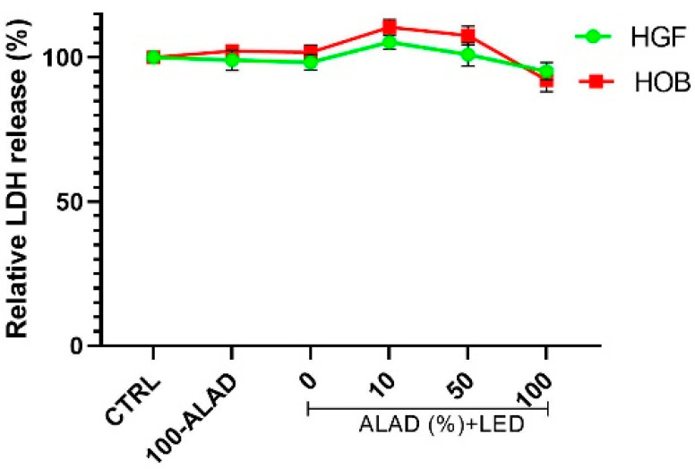
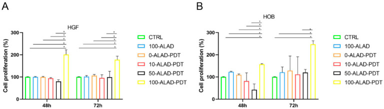
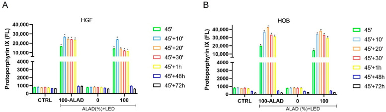
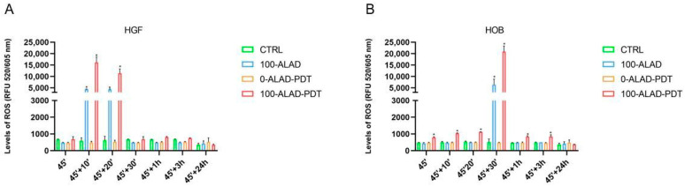
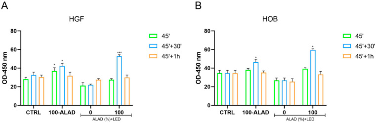
This study aimed to evaluate the effects of a new photodynamic protocol (ALAD-PDT), consisting of 5% 5-aminolevulinic acid-gel and 630 nm-LED, already used for antibacterial effects in the treatment of periodontitis, on human gingival fibroblasts (HGF) and primary human osteoblasts (HOB). HGF and HOB were incubated with different ALAD concentrations for 45 min, and subsequently irradiated with 630 nm-LED for 7 min. Firstly, the cytotoxicity at 24 h and proliferation at 48 and 72 h were assessed. Then the intracellular content of the protoporphyrin IX (PpIX) of the ROS and the superoxide dismutase (SOD) activity were investigated at different times. Each result was compared with untreated and unirradiated cells as the control. Viable and metabolic active cells were revealed at any concentrations of ALAD-PDT, but only 100-ALAD-PDT significantly enhanced the proliferation rate. The PpIX fluorescence significantly increased after the addition of 100-ALAD, and decreased after the irradiation. Higher ROS generation was detected at 10 min in HGF, and at 30 min in HOB. The activity of the SOD enzyme augmented at 30 min in both cell types. In conclusion, ALAD-PDT not only showed no cytotoxic effects, but had pro-proliferative effects on HGF and HOB, probably via ROS generation.

Keywords: 5-delta aminolevulinic acid; periodontal tissues; photodynamic therapy; protoporphyrin; reactive oxygen species.


Conflict of interest statement

The authors declare no conflict of interest.

Figures

Figure 1
Figure 1
Cytotoxicity assay. HGF and HOB were subjected to ALAD-PDT treatment, and LDH release was measured at 24 h. The data are expressed as percentages with respect to the control (CTRL).
Figure 2
Figure 2
Cell morphology. HGF were subjected to ALAD-PDT protocol and stained after 24 h with toluidine blue. (A) CTRL, (B) 0-ALAD-PDT, (C) 100-ALAD, (D) 100-ALAD-PDT.
Figure 3
Figure 3
Cell staining. HOB were subjected to ALAD-PDT protocol and stained after 24 h with toluidine blue. (A) CTRL, (B) 0-ALAD-PDT, (C) 100-ALAD, (D) 100-ALAD-PDT.
Figure 4
Figure 4
Cell proliferation assay. HGF (A) and HOB (B)were treated with different concentrations of ALAD gel and exposed to 630 nm LED. Cell growth was measured after 48 h and 72 h. Data are expressed in percentages with respect to control (CTRL). Data are presented as mean ± SD (error bars) of three independent experiments. The proliferation rate was significantly higher with the complete treatment 100-ALAD-PDT at 48 h for HGF, and 72 h for HOB. (* p < 0.0001).
Figure 5
Figure 5
Levels of intracellular PpIX. Fluorescence of PpIX was measured (λex = 405 nm; λem = 608 nm) immediately after the end of ALAD-PDT (45′) and after 10 min (45′ + 10′), 20 min (45′ + 20′), 30 min (45′ + 30′), 1 h (45′ + 1 h), 48 h (45′ + 48 h) and 72 h (45′ + 72 h). Data are presented as mean ± SD of three independent experiments. The statistical analysis was performed by ANOVA test comparing each value to its control (* p < 0.0001). A peak of PpIX fluorescence was detected at 10′ for HGF (A) and at 20′ for HOB (B).
Figure 6
Figure 6
Levels of ROS. The quantification of ROS was measured after the ALAD-PDT protocol at the end of the treatment (45′), at 10 min (45′ + 10′), at 20 min (45′ + 20′), at 30 min (45′ + 30′), at 1 h (45′ + 1 h), at 3 h (45′ + 3 h), and at 24 h (45′ + 24 h). Data are reported as mean ± SD of three independent experiments and expressed in the relative fluorescent unit (RFU), measured at λex/em 520/605 nm. The largest increment was observed after 10′ for HGF (A) and 30′ for HOB (B). The statistical analysis was performed by ANOVA test comparing each value to its control (* p < 0.0001).
Figure 7
Figure 7
SOD activity. Levels of SOD were detected after the application of ALAD-PDT protocol (45′) and after 30′ and 1 h from its end. Data are expressed as mean ± SD and measured as optical density (OD). A higher SOD activity was observed in ALAD-PDT treated cells after 30′ for both cell lines. The statistical analysis was performed by ANOVA test comparing each value to its control (* p < 0.05; *** p < 0.0001).

Similar articles

Cited by

References

    1. D’ercole S., di Lodovico S., Iezzi G., Pierfelice T.V., D’amico E., Cipollina A., Piattelli A., Cellini L., Petrini M. Complex Electromagnetic Fields Reduce Candida Albicans Planktonic Growth and Its Adhesion to Titanium Surfaces. Biomedicines. 2021;9:1261. doi: 10.3390/biomedicines9091261. - DOI - PMC - PubMed
    1. Petrini M., di Lodovico S., Iezzi G., Cipollina A., Piattelli A., Cellini L., D’Ercole S. Effects of Complex Electromagnetic Fields on Candida Albicans Adhesion and Proliferation on Polyacrylic Resin. Appl. Sci. 2021;11:6786. doi: 10.3390/app11156786. - DOI - PubMed
    1. Petrini M., Trentini P., Tripodi D., Spoto G., D’Ercole S. In Vitro Antimicrobial Activity of LED Irradiation on Pseudomonas Aeruginosa. J. Photochem. Photobiol. B. 2017;168:25–29. doi: 10.1016/j.jphotobiol.2017.01.020. - DOI - PubMed
    1. Gwynne P.J., Gallagher M.P. Light as a Broad-Spectrum Antimicrobial. Front. Microbiol. 2018;9:119. doi: 10.3389/fmicb.2018.00119. - DOI - PMC - PubMed
    1. D’Ercole S., Spoto G., Trentini P., Tripodi D., Petrini M. In Vitro Inactivation of Enterococcus Faecalis with a Led Device. J. Photochem. Photobiol. B. 2016;160:172–177. doi: 10.1016/j.jphotobiol.2016.04.015. - DOI - PubMed

LinkOut - more resources