

Are Columnar Cell Lesions the Earliest Non-Obligate Precursor in the Low-Grade Breast Neoplasia Pathway?
- PMID: 36005185
- PMCID: PMC9406596
- DOI: 10.3390/curroncol29080447
Are Columnar Cell Lesions the Earliest Non-Obligate Precursor in the Low-Grade Breast Neoplasia Pathway?
Abstract
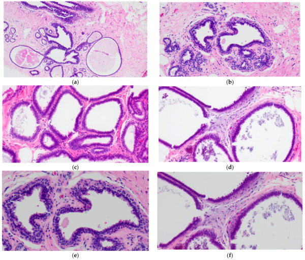
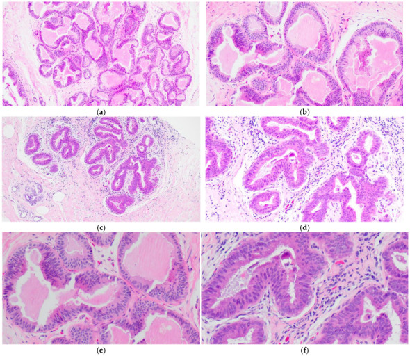
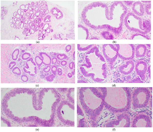
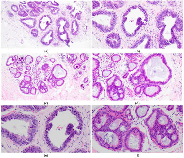
Columnar cell lesions (CCLs) of the breast comprise a spectrum of morphologic alterations of the terminal duct lobular unit involving variably dilated and enlarged acini lined by columnar epithelial cells. The World Health Organization currently classifies CCLs without atypia as columnar cell change (CCC) and columnar cell hyperplasia (CCH), whereas flat epithelial atypia (FEA) is a unifying term encompassing both CCC and CCH with cytologic atypia. CCLs have been increasingly recognized in stereotactic core needle biopsies (CNBs) performed for the assessment of calcifications. CCLs are believed to represent the earliest non-obligate precursor of low-grade invasive breast carcinomas as they share molecular alterations and often coexist with entities in the low-grade breast neoplasia pathway. Despite this association, however, the risk of progression of CCLs to invasive breast carcinoma appears low and may not exceed that of concurrent proliferative lesions. As the reported upgrade rates of pure CCL/FEA when identified as the most advanced high-risk lesion on CNB vary widely, the management of FEA diagnosed on CNB remains controversial. This review will include a historical overview of CCLs and will examine histologic diagnostic criteria, molecular alterations, prognosis and issues related to upgrade rates and clinical management.
Keywords: breast; carcinogenesis; columnar cell lesions; low-grade neoplasia; precursor.
Conflict of interest statement
The authors declare no conflict of interest.
Figures
Similar articles
-
Dimorphic cells: a common feature throughout the low nuclear grade breast neoplasia spectrum.Virchows Arch. 2023 Feb;482(2):369-375. doi: 10.1007/s00428-022-03438-w. Epub 2022 Nov 15. Virchows Arch. 2023. PMID: 36378325 Free PMC article.
-
Proliferating activity in columnar cell lesions of the breast.Virchows Arch. 2006 Dec;449(6):617-21. doi: 10.1007/s00428-006-0296-0. Epub 2006 Oct 6. Virchows Arch. 2006. PMID: 17024423
-
Are columnar cell lesions the earliest histologically detectable non-obligate precursor of breast cancer?Virchows Arch. 2008 Jun;452(6):589-98. doi: 10.1007/s00428-008-0609-6. Epub 2008 Apr 24. Virchows Arch. 2008. PMID: 18437416 Review.
-
Assessment of "grading" with Ki-67 and c-kit immunohistochemical expressions may be a helpful tool in management of patients with flat epithelial atypia (FEA) and columnar cell lesions (CCLs) on core breast biopsy.J Cell Physiol. 2009 Nov;221(2):343-9. doi: 10.1002/jcp.21858. J Cell Physiol. 2009. PMID: 19585492
-
Columnar cell lesions of the breast: an update and significance on core biopsy.Pathology. 2009 Jan;41(1):18-27. doi: 10.1080/00313020802563486. Pathology. 2009. PMID: 19089736 Review.
Cited by
-
Canine, Feline, and Murine Mammary Tumors as a Model for Translational Research in Breast Cancer.Vet Sci. 2025 Feb 19;12(2):189. doi: 10.3390/vetsci12020189. Vet Sci. 2025. PMID: 40005948 Free PMC article. Review.
-
[B3 lesions of the breast: histological, clinical, and epidemiological aspects : Update].Pathologie (Heidelb). 2023 Feb;44(1):5-16. doi: 10.1007/s00292-022-01180-3. Epub 2023 Jan 12. Pathologie (Heidelb). 2023. PMID: 36635403 Free PMC article. Review. German.
References
-
- Hoon Tan P.H., Ellis I., Allison K., Brogi E., Fox S.B., Lakhani S., Lazar A.J., Morris E.A., Sahin A., Salgado R., et al., editors. World Health Organization Classification of Tumours of the Breast. 5th ed. IARC Press; Lyon, France: 2019. - PubMed
-
- Luiten J.D., Korte B., Voogd A.C., Vreuls W., Luiten E.J.T., Strobbe L.J., Rutten M.J.C.M., Plaisier M.L., Lohle P.N., Hooijen M.J.H., et al. Trends in Frequency and Outcome of High-Risk Breast Lesions at Core Needle Biopsy in Women Recalled at Biennial Screening Mammography, a Multiinstitutional Study. Int. J. Cancer. 2019;145:2720–2727. doi: 10.1002/ijc.32353. - DOI - PMC - PubMed
Publication types
MeSH terms
LinkOut - more resources
Full Text Sources
Medical
Research Materials
