

Fulminant Influenza A Myocarditis Complicated by Transient Ventricular Wall Thickening and Cardiac Tamponade
- PMID: 36005267
- PMCID: PMC9408225
- DOI: 10.3390/idr14040065
Fulminant Influenza A Myocarditis Complicated by Transient Ventricular Wall Thickening and Cardiac Tamponade
Abstract
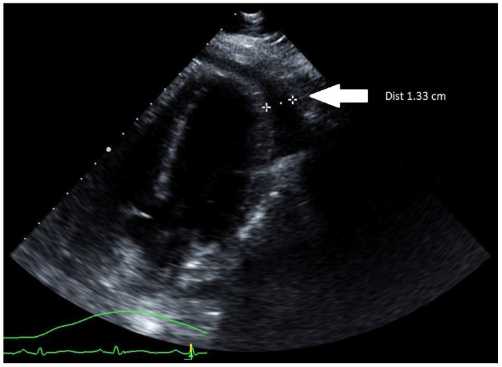
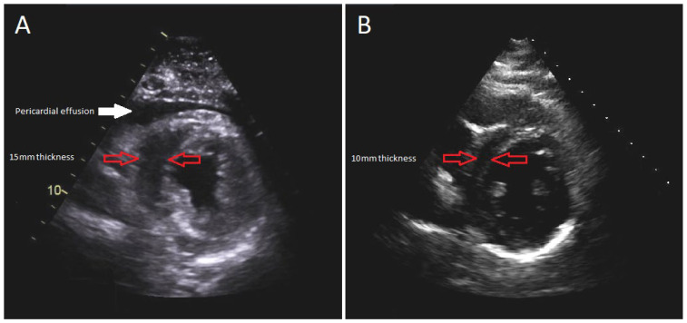
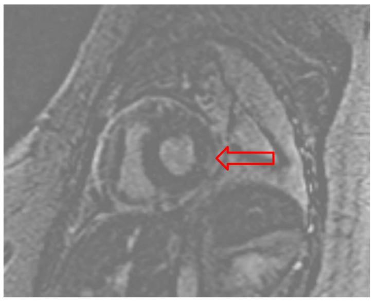
Myocarditis is an infrequent complication of influenza infection that is most often diagnosed clinically in the setting of confirmed influenza infection and elevated cardiac enzymes. Pericarditis can also occur in cases of influenza myocarditis and may require pericardiocentesis for tamponade. Patients with fulminant myocarditis have cardiogenic shock; however, echocardiographic findings may be subtle, showing a preserved ejection fraction and diffuse left ventricular wall thickening (compared to baseline) due to inflammatory edema. Recognizing these echocardiographic findings in the appropriate clinical setting facilitates the early recognition of fulminant myocarditis. Therefore, we report a case of fulminant influenza A myocarditis in healthy 37-year-old women complicated by transient left ventricular wall thickening and tamponade, highlighting the importance of early diagnosis and supportive management for a successful outcome.
Keywords: cardiac tamponade; fulminant myocarditis; influenza A; myocardial edema.
Conflict of interest statement
The authors declare no conflict of interest.
Figures
Similar articles
-
Fulminant pH1N1-09 influenza-associated myocarditis in pediatric patients.Pediatr Crit Care Med. 2011 Mar;12(2):e99-e101. doi: 10.1097/PCC.0b013e3181e28887. Pediatr Crit Care Med. 2011. PMID: 20601924 Free PMC article.
-
[Acute viral myocarditis with transient concentric pseudohypertrophy of the left ventricle complicated by cardiogenic shock].G Ital Cardiol. 1993 Dec;23(12):1223-8. G Ital Cardiol. 1993. PMID: 8174874 Italian.
-
Hemodynamics of cardiac tamponade during extracorporeal membrane oxygenation support in a patient with fulminant myocarditis.J Cardiol Cases. 2018 Oct 25;19(1):22-24. doi: 10.1016/j.jccase.2018.08.009. eCollection 2019 Jan. J Cardiol Cases. 2018. PMID: 30693054 Free PMC article.
-
Influenza Myopericarditis and Pericarditis: A Literature Review.J Clin Med. 2022 Jul 15;11(14):4123. doi: 10.3390/jcm11144123. J Clin Med. 2022. PMID: 35887887 Free PMC article. Review.
-
Transient ventricular wall thickening in acute myocarditis: a serial echocardiographic and histopathologic study.Jpn Circ J. 2001 Oct;65(10):863-6. doi: 10.1253/jcj.65.863. Jpn Circ J. 2001. PMID: 11665789 Review.
Cited by
-
Heavy hearts: the impact of influenza on young lives, a rare case report.Iran J Microbiol. 2025 Jun;17(3):511-515. doi: 10.18502/ijm.v17i3.18834. Iran J Microbiol. 2025. PMID: 40612725 Free PMC article.
References
Publication types
LinkOut - more resources
Full Text Sources
