. 2022 Oct 26;13(5):e0144022.
doi: 10.1128/mbio.01440-22. Epub 2022 Aug 25.

Toxin-Producing Endosymbionts Shield Pathogenic Fungus against Micropredators

Affiliations

Toxin-Producing Endosymbionts Shield Pathogenic Fungus against Micropredators

Ingrid Richter et al. mBio. .

Abstract

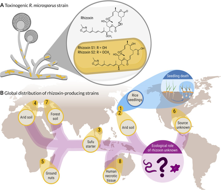
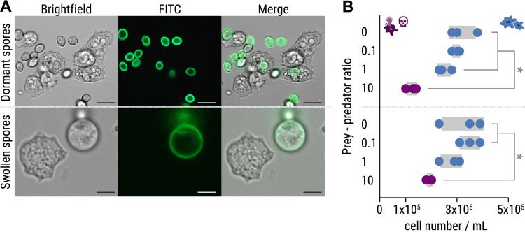
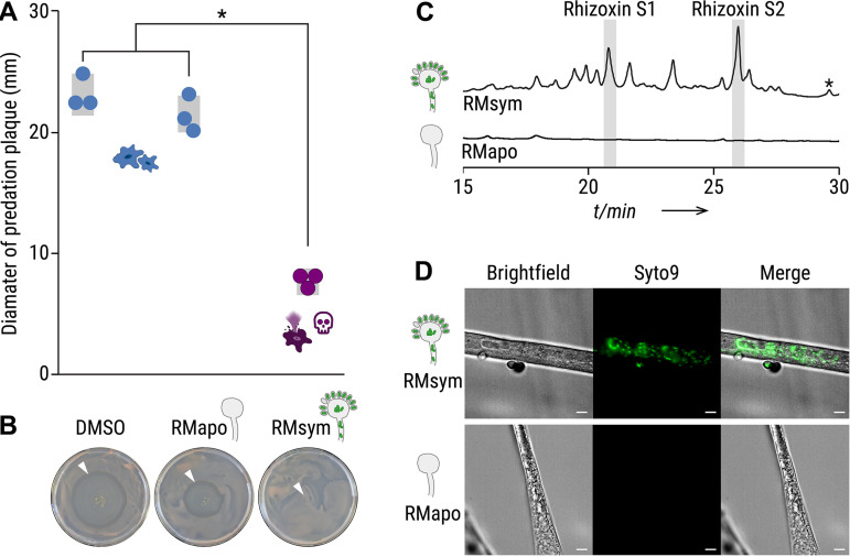
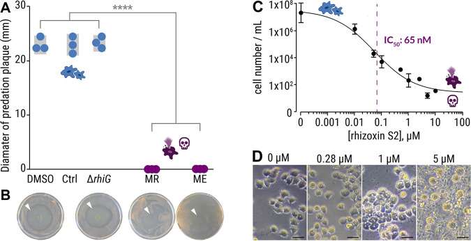
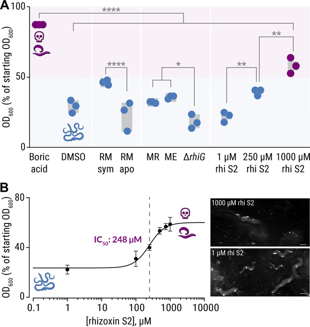
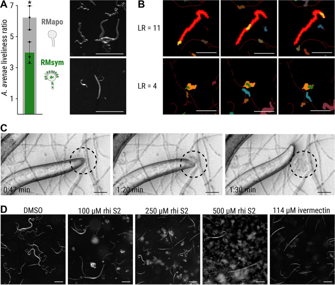
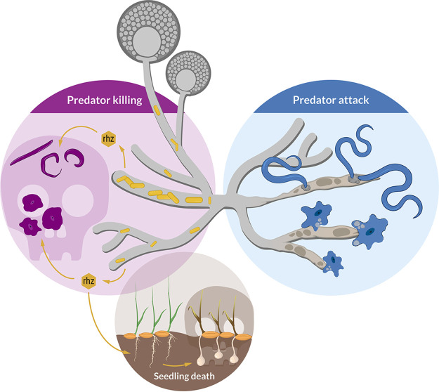
The fungus Rhizopus microsporus harbors a bacterial endosymbiont (Mycetohabitans rhizoxinica) for the production of the antimitotic toxin rhizoxin. Although rhizoxin is the causative agent of rice seedling blight, the toxinogenic bacterial-fungal alliance is, not restricted to the plant disease. It has been detected in numerous environmental isolates from geographically distinct sites covering all five continents, thus raising questions regarding the ecological role of rhizoxin beyond rice seedling blight. Here, we show that rhizoxin serves the fungal host in fending off protozoan and metazoan predators. Fluorescence microscopy and coculture experiments with the fungivorous amoeba Protostelium aurantium revealed that ingestion of R. microsporus spores is toxic to P. aurantium. This amoebicidal effect is caused by the dominant bacterial rhizoxin congener rhizoxin S2, which is also lethal toward the model nematode Caenorhabditis elegans. By combining stereomicroscopy, automated image analysis, and quantification of nematode movement, we show that the fungivorous nematode Aphelenchus avenae actively feeds on R. microsporus that is lacking endosymbionts, whereas worms coincubated with symbiotic R. microsporus are significantly less lively. This study uncovers an unexpected ecological role of rhizoxin as shield against micropredators. This finding suggests that predators may function as an evolutionary driving force to maintain toxin-producing endosymbionts in nonpathogenic fungi. IMPORTANCE The soil community is a complex system characterized by predator-prey interactions. Fungi have developed effective strategies to defend themselves against predators. Understanding these strategies is of critical importance for ecology, medicine, and biotechnology. In this study, we shed light on the defense mechanisms of the phytopathogenic Rhizopus-Mycetohabitans symbiosis that has spread worldwide. We report an unexpected role of rhizoxin, a secondary metabolite produced by the bacterium M. rhizoxinica residing within the hyphae of R. microsporus. We show that this bacterial secondary metabolite is utilized by the fungal host to successfully fend off fungivorous protozoan and metazoan predators and thus identified a fundamentally new function of this infamous cytotoxic compound. This endosymbiont-dependent predator defense illustrates an unusual strategy employed by fungi that has broader implications, since it may serve as a model for understanding how animal predation acts as an evolutionary driving force to maintain endosymbionts in nonpathogenic fungi.

Keywords: Rhizopus; microbial ecology; microbial interactions; natural products; rhizoxin; secondary metabolism; symbiosis.


Conflict of interest statement

The authors declare no conflict of interest.

Figures

FIG 1
FIG 1
Global distribution of a toxin-producing bacterial-fungal symbiosis. (A) Symbiotic bacteria (Mycetohabitans sp.) residing within the fungal hypha of R. microsporus, produce a mixture of toxic secondary metabolites (rhizoxins). (B) Rhizoxin-producing Rhizopus-Mycetohabitans strains were isolated from environmental samples from geographically distinct sites covering all five continents. In one of the eight toxinogenic strains (R. microsporus ATCC 62417, blue), rhizoxin causes blight disease in rice seedlings, while the ecological role of rhizoxin in the other, nonpathogenic Rhizopus strains is currently unknown.
FIG 2
FIG 2
Predation of Protostelium aurantium on spores of R. microsporus. (A) Fluorescence microscopy images showing FITC-stained, dormant R. microsporus spores (top) and ingestion of a swollen R. microsporus spore by P. aurantium (bottom). Scale bars, 5 μm. (B) Feeding of P. aurantium on dormant spores (top) leads to a reduced survival rate of P. aurantium compared to swollen spores (bottom). n = 3 independent replicated experiments ± 1 SEM. One-way ANOVA was performed with Tukey’s multiple-comparison test (*, P < 0.05; see also Table S1B).
FIG 3
FIG 3
Culture extracts from symbiotic Rhizopus microsporus kills Protostelium aurantium. (A) The survival of P. aurantium, indicated by the diameter of the predation plaque (clearance of yeast), is significantly reduced in cultures that were exposed to 2% crude culture extract from symbiotic R. microsporus (RMsym). Incubation with solvent alone (DMSO) or apo-symbiotic R. microsporus (RMapo) has no effect on the viability of P. aurantium. Circles indicate independent replicated experiments (n = 3) ± 1 SEM (gray bars). One-way ANOVA with Tukey’s multiple-comparison test was performed (*, P < 0.0001; see Table S2). (B) Photographs of yeast agar plates showing the predation plaque by P. aurantium (arrowheads). (C) HPLC profiles of crude extracts from symbiotic and endosymbiont-free R. microsporus showing a mixture of rhizoxin derivatives, including the two major bacterial rhizoxin congeners (rhizoxin S1 and rhizoxin S2). The peak correlating to rhizoxin is marked with an asterisk (*). Monitoring was done at 310 nm (see Fig. S2). The peak areas were integrated to calculate the concentration of rhizoxin S1 (1.2 μM) and rhizoxin S2 (1.7 μM), as well as all rhizoxin congeners combined (8.7 μM). (D) Fluorescence microscopy images of symbiotic R. microsporus and endosymbiont-free R. microsporus. Green fluorescence indicates presence of endosymbionts (SYTO9). Scale bars, 5 μm.
FIG 4
FIG 4
Culture extracts from axenic Mycetohabitans sp. kill Protostelium aurantium. (A) The viability of P. aurantium, indicated by the diameter of the predation plaque (clearance of yeast), is significantly reduced in cultures that were exposed to 2% crude culture extract from axenically grown endosymbiotic M. rhizoxinica HKI-0454 (labeled MR) or M. endofungorum HKI-0456 (labeled ME). Incubation with solvent alone (DMSO), extract from culture medium (Ctrl), or rhizoxin-deficient M. rhizoxinicarhiG) has no effect on the viability of P. aurantium. Circles indicate independent replicated experiments (n = 3) ± 1 SEM (gray bars). One-way ANOVA with Tukey’s multiple-comparison test was performed (****, P < 0.0001; see Table S3). (B) Photographs of yeast agar plates showing the predation plaque by P. aurantium (arrowheads). (C) Liquid survival assay of P. aurantium supplemented with the bacterial rhizoxin S2. Data points represent three independent replicated experiments (n = 3) ± 1 SEM. (D) Microscopic images showing the growth of P. aurantium in the presence of rhizoxin S2. Scale bars, 20 μm.
FIG 5
FIG 5
Inhibitory effects of crude extracts and pure rhizoxin S2 on C. elegans. (A) C. elegans, coincubated with E. coli OP50 cells as food source, were exposed to 2% crude culture extracts from symbiotic R. microsporus (RMsym), endosymbiont-free Rhizopus microsporus (RMapo), axenically grown endosymbiotic M. rhizoxinica HKI-0454 (labeled MR), Mycetohabitans endofungorum HKI-0456 (labeled ME), and rhizoxin-deficient M. rhizoxinicarhiG), as well as pure rhizoxin S2 (rhi S2). Since the number of viable nematode worms in the suspension is directly related to the E. coli cell density, the OD600 values were plotted as a percentage of the starting OD600. Incubation with 18 mM boric acid (positive control) kills most of the nematodes (E. coli density of 80%), while exposure to crude culture extracts has a mild effect on C. elegans viability. Circles indicate independent replicated experiments (n = 3) ± 1 SEM (gray bars). One-way ANOVA with Tukey’s multiple-comparison test was performed (*, P < 0.03; **, P < 0.002; ****, P < 0.0001; see Table S4). (B) Liquid feeding inhibition assay of C. elegans supplemented with the bacterial rhizoxin S2. Data points represent three independent replicated experiments (n = 3) ± 1 SEM. Microscopic images of nematodes exposed to pure rhizoxin S2 are shown. Scale bars, 200 μm.
FIG 6
FIG 6
Feeding inhibition of A. avenae on R. microsporus. (A) A. avenae was coincubated with symbiotic R. microsporus (RMsym) or endosymbiont-free R. microsporus (RMapo) for 2 to 3 weeks. Nematode movement was recorded using a stereomicroscope with a frame rate of 1 fps. The liveliness of the worms was calculated from the ratio of the area covered by a worm, divided by the area of the worm itself, and scaled to the full length of the movie. The minimum scaled liveliness ratio (LR) for a live worm was set to 1.5, below this value the worm was declared inactive/dead. n = 3 independent replicated experiments ± 1 SEM. An unpaired t test with Welch’s correction was performed (*, P < 0.05; see Table S5). Microscope images of A. avenae used for analysis. Scale bars, 500 μm. (B) Illustrations of the LR at high (top) and medium (bottom) values. (Top) The worm shown in orange covers the red footprint area during the time course of the experiment. These images show the first (left column), middle (middle column), and final (right column) time points of the movie. The activity of a worm was characterized by dividing the endpoint footprint by the area of the worm at each time point. The resulting LR was 11.5 for the worm in the top row, thus indicating a very active nematode. (Bottom) A less active worm (green area) covered a smaller footprint (orange area), as shown by the LR value of 4.0. Scale bars, 300 μm. See the live videos of the segmented worms and their footprints in Videos S11 and S12 (https://doi.org/10.5281/zenodo.6827988) for the worms with LR = 11.5 and LR = 4.0, respectively. (C) Time-lapse images of A. avenae feeding on endosymbiont-free R. microsporus (black circle). Endosymbiont-free R. microsporus ATCC 62417/S was coincubated with A. avenae for 24 h in a microchannel slide (Ibidi), and feeding was recorded on a spinning disc microscope (see Movie S1). Scale bars, 20 μm. No feeding was observed in worms that were coincubated with symbiotic R. microsporus (see Fig. S1D; see also Video S2 [https://doi.org/10.5281/zenodo.6827988]). (D) Microscopic images of A. avenae after exposure to different concentrations of pure rhizoxin S2 (rhi S2). Worms were healthy and alive when exposed to the solvent control (DMSO). Exposure to 114 μM ivermectin killed all worms. See the live videos of nematode movement in Videos S3 to S7 (https://doi.org/10.5281/zenodo.6827988). Scale bars, 200 μm.
FIG 7
FIG 7
Schematic model of the ecological role of rhizoxin-producing endofungal bacteria (M. rhizoxinica). The fungal host (Rhizopus microsporus) utilizes the bacterial secondary metabolite rhizoxin to fend off fungivorous micropredators such as amoeba and nematodes. The absence of endofungal bacteria leads to R. microsporus being attacked and subsequently killed by protozoan and metazoan predators. The establishment of the Rhizopus-Mycetohabitans symbiosis may have first developed to provide protection against fungal predators, with the emergence of plant pathogenicity developing later.

Similar articles

Cited by

References

    1. Lackner G, Partida-Martinez LP, Hertweck C. 2009. Endofungal bacteria as producers of mycotoxins. Trends Microbiol 17:570–576. doi:10.1016/j.tim.2009.09.003. - DOI - PubMed
    1. Scherlach K, Graupner K, Hertweck C. 2013. Molecular bacteria-fungi interactions: effects on environment, food, and medicine. Annu Rev Microbiol 67:375–397. doi:10.1146/annurev-micro-092412-155702. - DOI - PubMed
    1. Lackner G, Hertweck C. 2011. Impact of endofungal bacteria on infection biology, food safety, and drug development. PLoS Pathog 7:e1002096. doi:10.1371/journal.ppat.1002096. - DOI - PMC - PubMed
    1. Iwasaki S, Kobayashi H, Furukawa J, Namikoshi M, Okuda S, Sato Z, Matsuda I, Noda T. 1984. Studies on macrocyclic lactone antibiotics. VII. Structure of a phytotoxin “rhizoxin” produced by Rhizopus chinensis. J Antibiot (Tokyo) 37:354–362. doi:10.7164/antibiotics.37.354. - DOI - PubMed
    1. Partida-Martinez LP, Hertweck C. 2005. Pathogenic fungus harbors endosymbiotic bacteria for toxin production. Nature 437:884–888. doi:10.1038/nature03997. - DOI - PubMed

Publication types

LinkOut - more resources