. 2022 Jul 30;20(8):495.
doi: 10.3390/md20080495.

A Novel Marine Pyran-Isoindolone Compound Enhances Fibrin Lysis Mediated by Single-Chain Urokinase-Type Plasminogen Activator

Affiliations

A Novel Marine Pyran-Isoindolone Compound Enhances Fibrin Lysis Mediated by Single-Chain Urokinase-Type Plasminogen Activator

Chunli Gao et al. Mar Drugs. .

Abstract

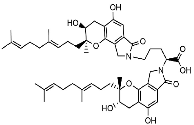
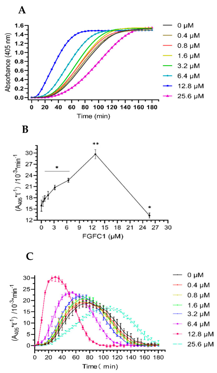
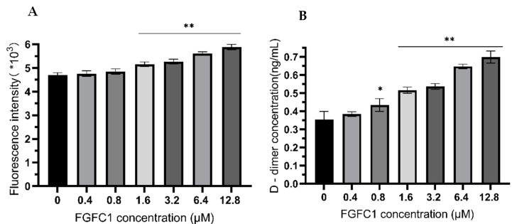
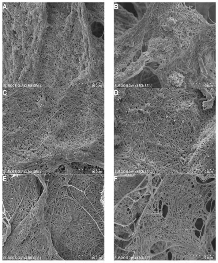
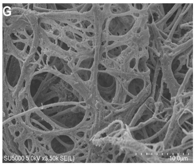
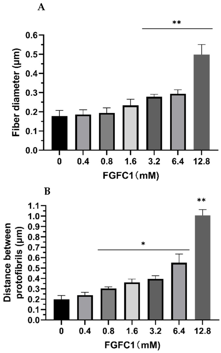
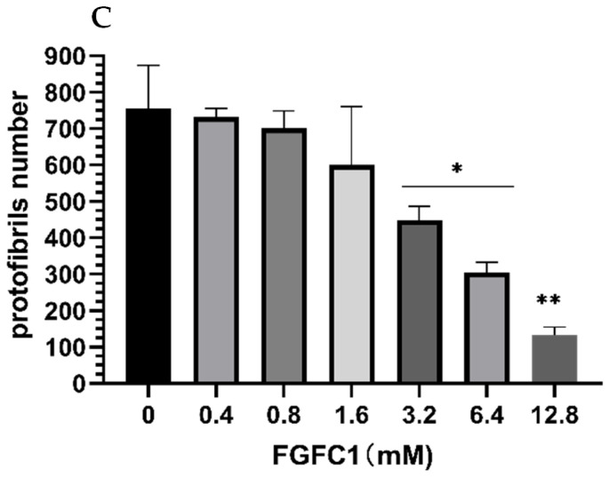
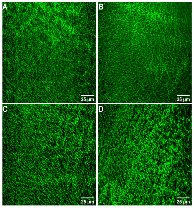
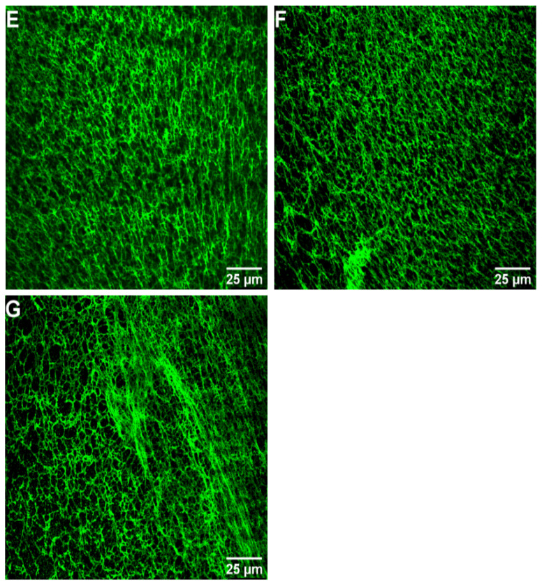
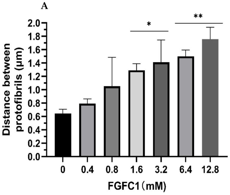
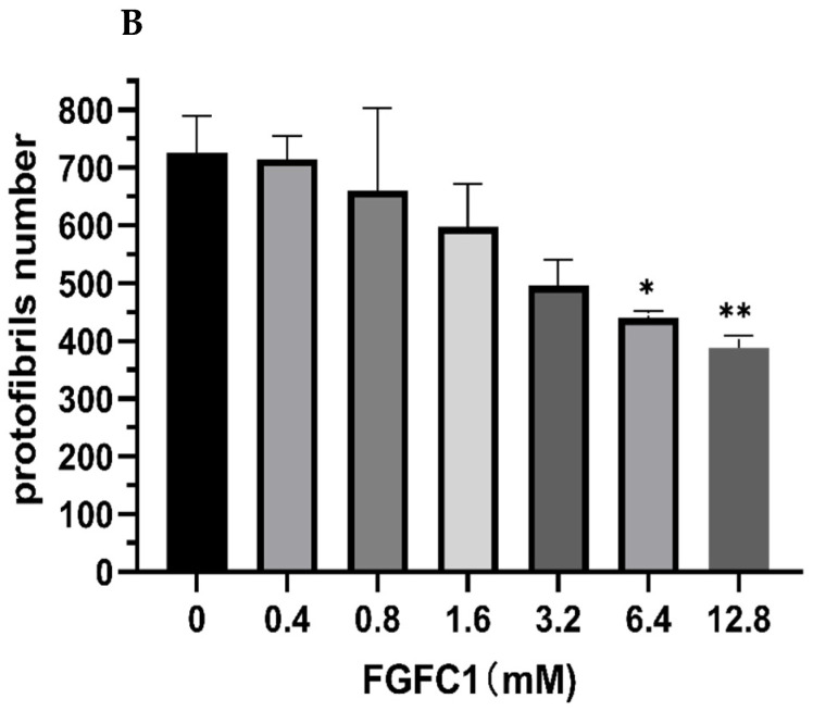
Fungi fibrinolytic compound 1 (FGFC1) is a rare pyran-isoindolone derivative with fibrinolytic activity. The aim of this study was to further determine the effect of FGFC1 on fibrin clots lysis in vitro. We constructed a fibrinolytic system containing single-chain urokinase-type plasminogen activator (scu-PA) and plasminogen to measure the fibrinolytic activity of FGFC1 using the chromogenic substrate method. After FITC-fibrin was incubated with increasing concentrations of FGFC1, the changes in the fluorescence intensity and D-dimer in the lysate were measured using a fluorescence microplate reader. The fibrin clot structure induced by FGFC1 was observed and analyzed using a scanning electron microscope and laser confocal microscope. We found that the chromogenic reaction rate of the mixture system increased from (15.9 ± 1.51) × 10−3 min−1 in the control group to (29.7 ± 1.25) × 10−3 min−1 for 12.8 μM FGFC1(p < 0.01). FGFC1 also significantly increased the fluorescence intensity and d-dimer concentration in FITC fibrin lysate. Image analysis showed that FGFC1 significantly reduced the fiber density and increased the fiber diameter and the distance between protofibrils. These results show that FGFC1 can effectively promote fibrin lysis in vitro and may represent a novel candidate agent for thrombolytic therapy.

Keywords: FGFC1; fibrin clot; fibrinolysis; fibrinolytic activity; plasminogen.


Conflict of interest statement

The authors declare no conflict of interest.

Figures

Figure 1
Figure 1
The structure of FGFC1.
Figure 2
Figure 2
Fibrinolytic activity of FGFC1 (0–25.6 μM). (A) The absorbance–time curve of the mixture system was measured at 405 nm at 37 °C for 180 min using a microplate reader. (B) The Kn (the slope of each curve) values of FGFC1 (0–25.6 μM) in the reaction system. Results expressed as the mean ± SD of three parallel experiments. * p < 0.05, ** p < 0.01, compared to the control group (absence FGFC1). (C) The reaction rates of FGFC1 (0–25.6 μM) at different times (0–180 min).
Figure 3
Figure 3
FGFC1 (0–12.8 μM) promotes fibrinolysis in vitro. (A) Fluorescence intensity of the mixture system was measured at the excitation wavelength of 495 nm and the emission wavelength of 520 nm after incubation at 37 °C for 20 min. (B) Enzyme-linked immunosorbent assay was used to detect effect of FGFC1 (0–12.8 μM) on lysis of FITC-fibrin. Results expressed by the mean ± SD of three parallel experiments. * p < 0.05, ** p < 0.01, compared to the control group (absence FGFC1).
Figure 4
Figure 4
Scanning electron microscopy images of fibrin clots (all scale bars are 10 μm). Clotting was initiated in the (A) absence and (BG) presence (0.4–12.8 μM) of FGFC1 by 1 U/mL thrombin and 15 mM CaCl2.
Figure 4
Figure 4
Scanning electron microscopy images of fibrin clots (all scale bars are 10 μm). Clotting was initiated in the (A) absence and (BG) presence (0.4–12.8 μM) of FGFC1 by 1 U/mL thrombin and 15 mM CaCl2.
Figure 5
Figure 5
Effect of FGFC1 (0–12.8 μM) on the molecular structure of fibrin fibers (scanning electron microscopy). (A) Diameter of fibrin fiber. (B) Distance between protofibrils within fibrin fibers. (C) Number of protofibrils. Results expressed by the mean ± SD of three parallel experiments. * p < 0.05, ** p < 0.01, compared to the control group (absence FGFC1).
Figure 5
Figure 5
Effect of FGFC1 (0–12.8 μM) on the molecular structure of fibrin fibers (scanning electron microscopy). (A) Diameter of fibrin fiber. (B) Distance between protofibrils within fibrin fibers. (C) Number of protofibrils. Results expressed by the mean ± SD of three parallel experiments. * p < 0.05, ** p < 0.01, compared to the control group (absence FGFC1).
Figure 6
Figure 6
Confocal laser scanning microscopy images of fibrin networks (all scale bars are 25 μm). Clotting was initiated in the (A) absence and (BG) presence (0.4–12.8 μM) of FGFC1 by 0.6 U/mL thrombin and 8 mM CaCl2. Fibrin networks were stained with FITC.
Figure 6
Figure 6
Confocal laser scanning microscopy images of fibrin networks (all scale bars are 25 μm). Clotting was initiated in the (A) absence and (BG) presence (0.4–12.8 μM) of FGFC1 by 0.6 U/mL thrombin and 8 mM CaCl2. Fibrin networks were stained with FITC.
Figure 7
Figure 7
Effect of FGFC1 (0–12.8 μM) on the molecular structure of fibrin fibers by laser scanning confocal microscopy. (A) Distance between protofibrils within fibrin fibers. (B) Number of protofibrils. Results expressed by the mean ± SD; n = 3. * p < 0.05, ** p < 0.01, compared to the control group (absence of FGFC1).
Figure 7
Figure 7
Effect of FGFC1 (0–12.8 μM) on the molecular structure of fibrin fibers by laser scanning confocal microscopy. (A) Distance between protofibrils within fibrin fibers. (B) Number of protofibrils. Results expressed by the mean ± SD; n = 3. * p < 0.05, ** p < 0.01, compared to the control group (absence of FGFC1).
Figure 8
Figure 8
The chromatogram of FGFC1.

References

    1. Plow E.F., Wang Y.M., Simon D.I. The search for new antithrombotic mechanisms and therapies that may spare hemostasis. Blood. 2018;131:1899–1902. doi: 10.1182/blood-2017-10-784074. - DOI - PMC - PubMed
    1. Alagarsamy K.N., Mathan S., Yan W., Rafieerad A., Sekaran S., Manego H., Dhingra S. Carbon nanomaterials for cardiovascular theranostics: Promises and challenges. Bioact. Mater. 2021;6:2261–2280. doi: 10.1016/j.bioactmat.2020.12.030. - DOI - PMC - PubMed
    1. Xie Z.L., Tian Y.B., Lv X., Xiao X., Zhan M.M., Cheng K.i., Li S.Y., Liao C.Z. The selectivity and bioavailability improvement of novel oral anticoagulants: An overview. Eur. J. Med. Chem. 2018;146:299–317. doi: 10.1016/j.ejmech.2018.01.067. - DOI - PubMed
    1. Wang Y.N., Chen H., Sheng R.L., Fu Z., Fan J.T., Wu W.H., Tu Q.D., Guo R.H. Synthesis and Bioactivities of Marine Pyran-Isoindolone Derivatives as Potential Antithrombotic Agents. Mar. Drugs. 2021;19:218. doi: 10.3390/md19040218. - DOI - PMC - PubMed
    1. Benjamin E.J., Muntner P., Alonso A., Bittencourt M.S., Callaway C.W., Carson A.P., Chamberlain A.M., Chang A.R., Cheng S., Das S.R., et al. Heart Disease and Stroke Statistics—2019 Update: A Report From the American Heart Association. Circulation. 2019;139:E56–E528. doi: 10.1161/CIR.0000000000000659. - DOI - PubMed