Review
. 2022 Jul 29;12(8):709.
doi: 10.3390/metabo12080709.

Altered TDP-43 Structure and Function: Key Insights into Aberrant RNA, Mitochondrial, and Cellular and Systemic Metabolism in Amyotrophic Lateral Sclerosis

Affiliations
Review

Altered TDP-43 Structure and Function: Key Insights into Aberrant RNA, Mitochondrial, and Cellular and Systemic Metabolism in Amyotrophic Lateral Sclerosis

Leanne Jiang et al. Metabolites. .

Abstract

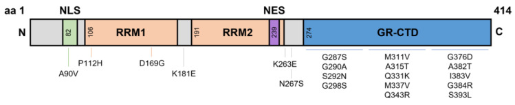
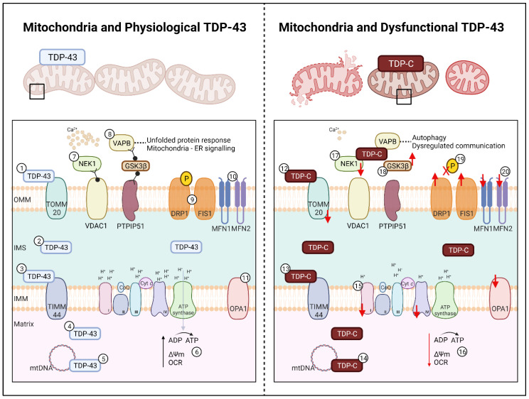
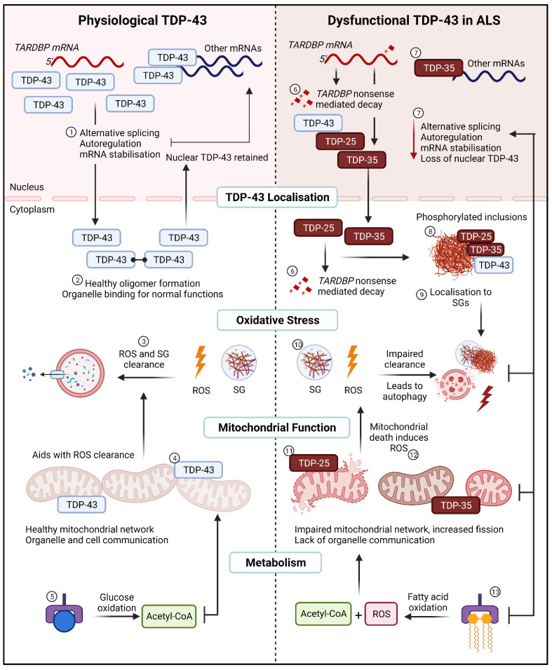
Amyotrophic lateral sclerosis (ALS) is a progressive and fatal neuromuscular disorder with no cure available and limited treatment options. ALS is a highly heterogeneous disease, whereby patients present with vastly different phenotypes. Despite this heterogeneity, over 97% of patients will exhibit pathological TAR-DNA binding protein-43 (TDP-43) cytoplasmic inclusions. TDP-43 is a ubiquitously expressed RNA binding protein with the capacity to bind over 6000 RNA and DNA targets-particularly those involved in RNA, mitochondrial, and lipid metabolism. Here, we review the unique structure and function of TDP-43 and its role in affecting the aforementioned metabolic processes in ALS. Considering evidence published specifically in TDP-43-relevant in vitro, in vivo, and ex vivo models we posit that TDP-43 acts in a positive feedback loop with mRNA transcription/translation, stress granules, cytoplasmic aggregates, and mitochondrial proteins causing a relentless cycle of disease-like pathology eventuating in neuronal toxicity. Given its undeniable presence in ALS pathology, TDP-43 presents as a promising target for mechanistic disease modelling and future therapeutic investigations.

Keywords: ALS; OXPHOS; RNA; TDP-43; amyotrophic lateral sclerosis; autoregulation; lipid metabolism; metabolism; mitochondria; mitochondrial dynamics; non-coding; splicing.


Conflict of interest statement

The authors declare no conflict of interest.

Figures

Figure 1
Figure 1
Domains of TDP-43 and known Single Nucleotide Polymorphism mutations. TDP-43 is a 43 kDa protein containing 414 amino acids. The NLS resides from aa82–98, with RRM1 and RRM2 residing at aa106–176 and aa191–259, respectively. The NES resides within RRM2 at aa239–250, and a GR-CTD is located at aa274–414. Most known single nucleotide polymorphisms reside on the GR-CTD. However, some mutations have been identified at the NLS and around the RRMs. aa, amino acid; NLS, nuclear localisation signal domain; RRM, RNA recognition motifs; NES, nuclear export signal; GR-CTD, glycine rich C-terminal domain.
Figure 2
Figure 2
Effect of TDP-43 on Mitochondria in ALS. Left: Mitochondria and Physiological TDP-43. In healthy cells length TDP-43 localises to (1) TOMM20 on the OMM, (2) within the IMS and to (3) TIMM44 of the IMM, (4) freely within the matrix, and (5) regulates mtDNA for OXPHOS complex expression. (6) OXPHOS complexes I-IV and ATP synthase facilitate ATP production, facilitating the maintenance of the membrane potential and a healthy oxygen consumption rate. (7) Mitochondrial proteins NEK1 and VDAC1 support calcium signalling. (8) The mitochondrial-endoplasmic reticulum complex, PTPIP51-GSK3β-VAPB ensures proper protein folding and organelle communication. The mitochondrial dynamics related proteins for (9) fission on the OMM (DRP1, FIS1) and (10) fusion on the OMM (MFN1, MFN2) and the (11) IMM (OPA1) facilitate mitochondrial network homeostasis and regulation. Right: Mitochondria and Dysfunctional TDP-43. In ALS, fragmented TDP of 25 kDa and 35 kDa (denoted as TDP-C) localises to (12) TOMM20 and (13) TIMM44 causing downregulation and loss of mitochondria. TDP-C also co-localise to (14) mtDNA causing fragmentation, leading to a (15) loss of OXPHOS complex I and IV. (16) Loss of complex regulation leads to a decrease in ATP production, driving a loss of membrane potential and oxygen consumption rate. (17) TDP-C leads to a decrease in NEK1, causing a loss of VDAC1 and calcium signalling. (18) Localisation of TDP-C to GSK3β causes the loss of mitochondrial-endoplasmic reticulum signalling resulting in protein folding dysfunction and loss of organelle communication, which in turn leads to autophagy. Further impacts of TDP-C results in (19) DRP1 de-phosphorylation leading to (20) increased fission and decreased fusion. Upregulation of FIS1 and downregulation of MFN1, MFN2, and OPA1 are also observed in TDP-43 models leading to increased fission and mitochondrial fragmentation and subsequent cellular death. TDP-C; TDP-43 C-terminal fragments, OMM; outer mitochondrial membrane, IMS; inter-membrane space, IMM; inner mitochondrial matrix, ATP; adenosine triphosphate, CoQ; Coenzyme Q; Cyt c; cytochrome c. Created with BioRender.com accessed on 12 July 2022.
Figure 3
Figure 3
The relentless TDP-43 positive feedback loop in ALS. (Left), Physiological TDP-43. (1) In healthy cells TDP-43 regulates normal RNA metabolic functions such as alternative splicing, autoregulation, and mRNA stabilisation. (2) TDP-43 oligomers are generated in the cytoplasm and cleared upon return to the nucleus. (3) Under normal oxidative stress conditions, ROS and SG are cleared by the aid of healthy mitochondria and a lack of mutant TDP-43 aggregating with SGs. (4) Full-length TDP-43 binds to the mitochondria to undergo normal functions and aids in organelle and cell communication. (5) Glucose as the main substrate for energy utilisation for ATP in mitochondria is utilised. (Right), Dysfunctional TDP-43 in ALS. (6) Dysfunctional TDP-43 transcribes and translates fragmented TDP-43, some fragmented are sent for nonsense mediated decay in the nucleus or the cytoplasm. (7) A loss of RNA metabolic functions is observed alongside a loss of nuclear TDP-43. (8) TDP-43 and TDP-43 fragments localise to the cytoplasm, where they are phosphorylated and form aggregates. (9) These aggregates lead to sequestration into SGs. (10) Impaired clearance of ROS and SG results in autophagy and cell death through a lack of apoptosis. (11) Mutant TDP-43 bind to mitochondria to impair the mitochondrial network, as well as mitochondrial signalling and function. (12) Lack of mitochondrial function creates a feedback loop with impaired oxidative stress and ROS clearance. (13) Switch from glycolysis to fatty acid oxidation induces ROS alongside Acetyl-CoA for substrate utilisation in mitochondria. SG; stress granules, ROS; reactive oxygen species. Created with BioRender.com accessed on 12 July 2022.

References

    1. Mitchell J.D., Borasio G.D. Amyotrophic lateral sclerosis. Lancet. 2007;369:2031–2041. doi: 10.1016/S0140-6736(07)60944-1. - DOI - PubMed
    1. Zufiria M., Gil-Bea F.J., Fernandez-Torron R., Poza J.J., Muñoz-Blanco J.L., Rojas-Garcia R., Riancho J., de Munain A.L. ALS: A bucket of genes, environment, metabolism and unknown ingredients. Prog. Neurobiol. 2016;142:104–129. doi: 10.1016/j.pneurobio.2016.05.004. - DOI - PubMed
    1. Arai T., Hasegawa M., Akiyama H., Ikeda K., Nonaka T., Mori H., Mann D., Tsuchiya K., Yoshida M., Hashizume Y. TDP-43 is a component of ubiquitin-positive tau-negative inclusions in frontotemporal lobar degeneration and amyotrophic lateral sclerosis. Biochem. Biophys. Res. Commun. 2006;351:602–611. doi: 10.1016/j.bbrc.2006.10.093. - DOI - PubMed
    1. Neumann M., Sampathu D.M., Kwong L.K., Truax A.C., Micsenyi M.C., Chou T.T., Bruce J., Schuck T., Grossman M., Clark C.M. Ubiquitinated TDP-43 in frontotemporal lobar degeneration and amyotrophic lateral sclerosis. Science. 2006;314:130–133. doi: 10.1126/science.1134108. - DOI - PubMed
    1. Leigh P., Whitwell H., Garofalo O., Buller J., Swash M., Martin J., Gallo J.-M., Weller R., Anderton B. Ubiquitin-immunoreactive intraneuronal inclusions in amyotrophic lateral sclerosis: Morphology, distribution, and specificity. Brain. 1991;114:775–788. doi: 10.1093/brain/114.2.775. - DOI - PubMed

LinkOut - more resources