. 2022 Aug 9;12(8):735.
doi: 10.3390/metabo12080735.

Nicotine Exposure during Rodent Pregnancy Alters the Composition of Maternal Gut Microbiota and Abundance of Maternal and Amniotic Short Chain Fatty Acids

Affiliations

Nicotine Exposure during Rodent Pregnancy Alters the Composition of Maternal Gut Microbiota and Abundance of Maternal and Amniotic Short Chain Fatty Acids

Jasenka Zubcevic et al. Metabolites. .

Abstract

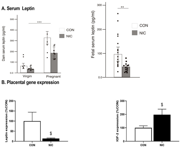
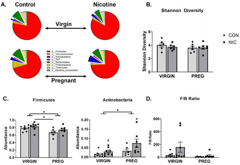
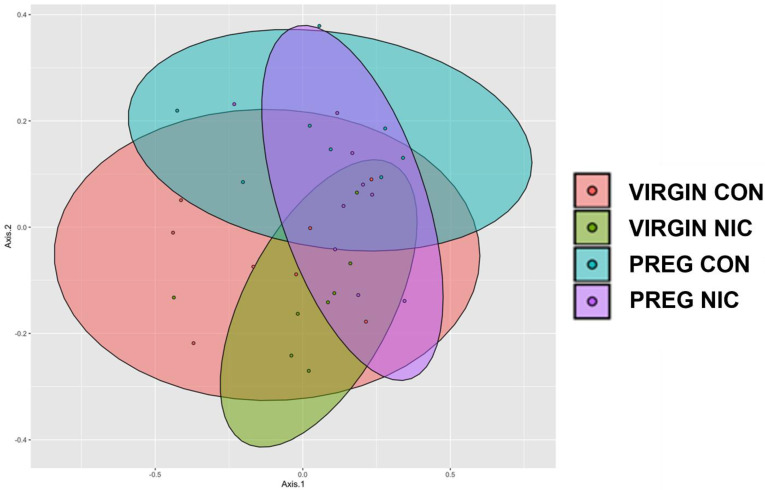
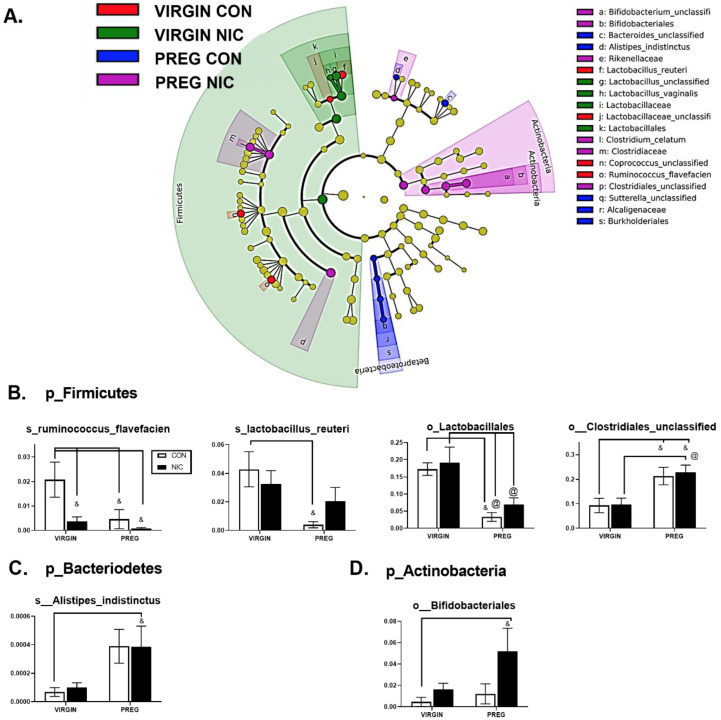
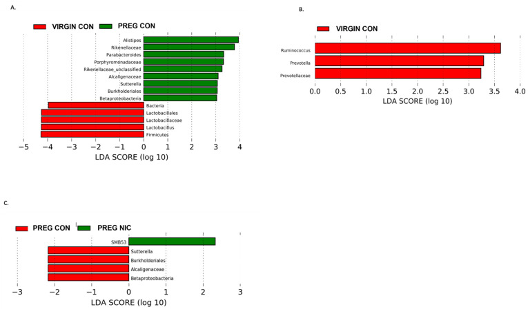
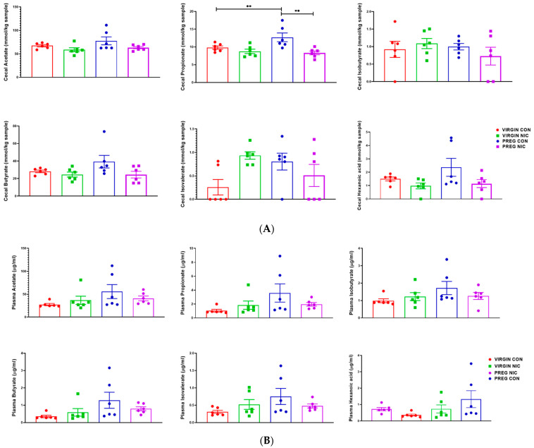
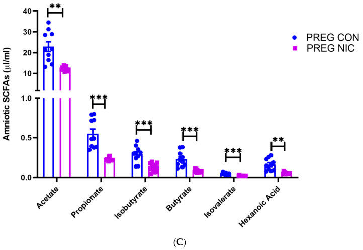
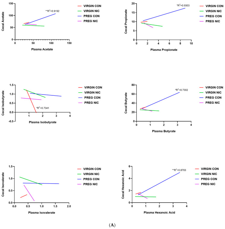
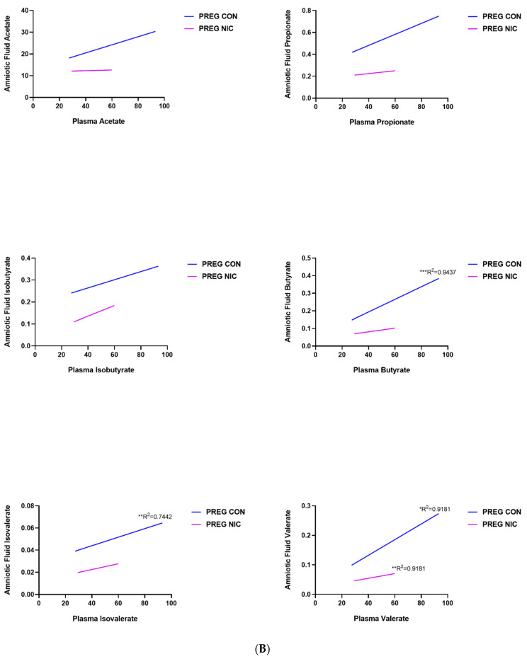
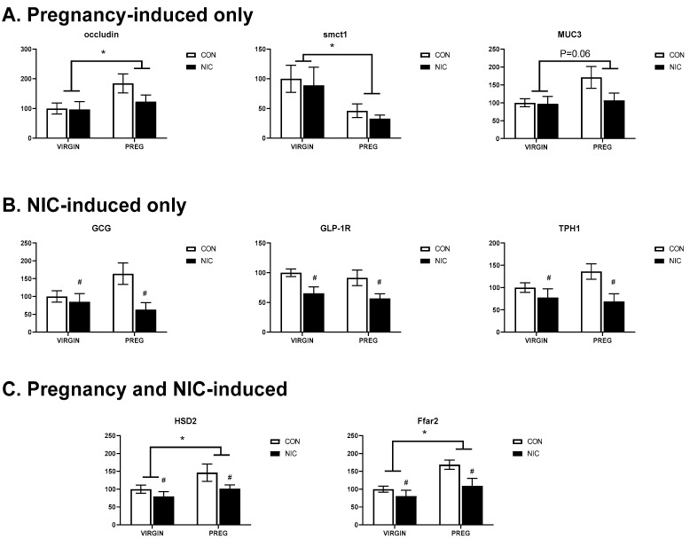
Tobacco smoking is the leading cause of preventable death. Numerous reports link smoking in pregnancy with serious adverse outcomes, such as miscarriage, stillbirth, prematurity, low birth weight, perinatal morbidity, and infant mortality. Corollaries of consuming nicotine in pregnancy, separate from smoking, are less explored, and the mechanisms of nicotine action on maternal-fetal communication are poorly understood. This study examined alterations in the maternal gut microbiome in response to nicotine exposure during pregnancy. We report that changes in the maternal gut microbiota milieu are an important intermediary that may mediate the prenatal nicotine exposure effects, affect gene expression, and alter fetal exposure to circulating short-chain fatty acids (SCFAs) and leptin during in utero development.

Keywords: SCFA; microbiota; pregnancy; smoking.


Conflict of interest statement

The authors declare no conflicts of interest.

Figures

Figure 1
Figure 1
Serum and placental leptin levels were reduced, and placental IGF-2 gene expression elevated following 13 days of nicotine exposure (NIC; 6 mg/kg/day) compared to controls (CON). (A) Serum leptin levels measured in adult female (left graph; n = 6–8/group) and fetuses (right graph; n = 12/group). (B) Placental gene expression for leptin (left graph) and IGF-2 (right graph; n = 8–11/group). Values are median ± SEM (A) and mean ± SEM (B); ** and *** indicate p < 0.01 and p < 0.001 respectively, between virgin and pregnant groups combined; # indicates p < 0.019 between NIC vs. CON in virgin and pregnant females combined (two-way ANOVA). $ indicates p < 0.05 between CON and NIC (Mann–Whitney test).
Figure 2
Figure 2
Nicotine (NIC) exposure shifts cecal bacterial abundance in virgin versus pregnant (preg; gestational day 19) Sprague Dawley rats. (A) Average proportion of bacteria within each phylum identified. (B) Gut microbial diversity as measured by Shannon diversity index. (C) Two-way ANOVA identified significant shifts in bacteria in phyla Firmicutes and Actinobacteria in pregnancy (p < 0.02). NIC also elevated the proportion of bacteria in p_Actinobacteria, independent of pregnancy status (p < 0.04). (D) The ratio of bacteria in p_Firmicutes to p_Bacteriodetes or F/B ratio was unchanged by pregnancy or NIC exposure. Values are mean ± SEM; Data were analyzed by two-way ANOVA with multiple comparisons. * (handle bars) indicates p < 0.02 between virgin and pregnant groups combined; # indicates p < 0.04 between NIC vs. CON in virgin and pregnant females combined; * (straight line) indicates p < 0.05 between treatment groups. n = 6–8 samples per group.
Figure 3
Figure 3
Principle coordinate analysis (PCoA) comparing the impact of nicotine exposure in virgin and pregnant female SD rats. NIC generally narrowed the clusters in both groups compared to CON, with the greatest shift during pregnancy. Data are based on 16s sequencing, n = 6–8 samples per group.
Figure 4
Figure 4
Nicotine exposure in rodent pregnancy induces selective changes in cecal bacterial abundance in Firmicutes, Bacteriodetes, and Actinobacteria phyla. (A) Taxonomic cladogram generated in Galaxy demonstrating shifts in taxonomic groups following nicotine exposure in the virgin versus pregnant rats (P). Each circle represents a bacterial taxon, with its diameter proportional to the taxon’s relative abundance. Yellow circles indicate no change. Colored circles indicate significant changes in abundance relative to other groups. Red—virgin controls; green—virgin nicotine exposed; blue—pregnant controls and purple—pregnant-nicotine exposed data. (BD) Two-way ANOVA analysis with multiple comparisons was performed and revealed significant differences in order (o_) or species (s_) between groups in the phylum Firmicutes, Bacteriodetes and Actinobactera, as annotated. Values are mean ± SEM; & indicates p < 0.05 between CON in virgin and other groups as indicated; @ indicates p < 0.02 between virgin nicotine versus other groups indicated. n = 6–8 samples per group.
Figure 5
Figure 5
Effect of pregnancy and nicotine on cecal bacterial abundance and composition in female SD rats. LDA analysis of cecal bacterial taxa demonstrates significant shifts in taxonomic groups during pregnancy alone (in (A) red = virgin control; green = pregnant control), during s.c. exposure to nicotine in female SD rats (in (B) red = virgin control, green = virgin nicotine), and during pregnancy with exposure to s.c. nicotine (in (C) red = pregnant control, green = pregnant nicotine) in SD dams. Each color represents a more abundant bacterial taxon relative to the other group. A marked decrease in Proteobacteria and specifically Betaproteobacteria in the PREG NIC vs. PREG CON group is shown (in (C)), while an elevated abundance in Betaproteobacteria may be important in normal rodent pregnancy (in (A)). Data are based on 16s sequencing analysis, n = 6–8 per group.
Figure 6
Figure 6
In (A), propionate increased during pregnancy in control (PREG CON), while nicotine (PREG NIC) exposure reduced cecal propionate levels in pregnant dams. All other cecal SCFAs were unchanged by pregnancy or NIC exposure. In (B), no significant differences were observed in circulating SCFAs between the groups. However, in (C), all measured SCFAs were significantly reduced in amniotic fluid collected from PREG NIC group. Values expressed as mean ± SEM; Data were analyzed using one-way ANOVA with multiple comparisons. ** p < 0.01, *** p < 0.001, n = 6–10 per group.
Figure 6
Figure 6
In (A), propionate increased during pregnancy in control (PREG CON), while nicotine (PREG NIC) exposure reduced cecal propionate levels in pregnant dams. All other cecal SCFAs were unchanged by pregnancy or NIC exposure. In (B), no significant differences were observed in circulating SCFAs between the groups. However, in (C), all measured SCFAs were significantly reduced in amniotic fluid collected from PREG NIC group. Values expressed as mean ± SEM; Data were analyzed using one-way ANOVA with multiple comparisons. ** p < 0.01, *** p < 0.001, n = 6–10 per group.
Figure 7
Figure 7
Relationship between cecal, plasma and amniotic fluid SCFAs in pregnant dams on nicotine. In (A), a positive correlation between cecal and circulating (plasma) acetate, propionate, butyrate and hexanoic acid was observed in pregnant control (PREG CON) dams alone. In (B), a positive correlation between circulating (plasma) and amniotic fluid levels of all measured SCFAs was observed in some samples from PREG CON dams. Linear regression analysis was performed, * p < 0.05, ** p < 0.01, *** p < 0.001 from zero; n = 8–10 samples per group.
Figure 7
Figure 7
Relationship between cecal, plasma and amniotic fluid SCFAs in pregnant dams on nicotine. In (A), a positive correlation between cecal and circulating (plasma) acetate, propionate, butyrate and hexanoic acid was observed in pregnant control (PREG CON) dams alone. In (B), a positive correlation between circulating (plasma) and amniotic fluid levels of all measured SCFAs was observed in some samples from PREG CON dams. Linear regression analysis was performed, * p < 0.05, ** p < 0.01, *** p < 0.001 from zero; n = 8–10 samples per group.
Figure 8
Figure 8
Pregnancy and NIC alter gene expression in cecal tissue. (A) Pregnancy (PREG) alone upregulates expression of occluding and mucin 3 (muc3) and downregulates expression of sodium-coupled monocarboxylate transporter 1 (smctl), the lactate transporter. (B) NIC downregulated expression of genes associated with glucagon (Gcg), glucagon-like protein type 1 receptor (Glp-1r) and tryptophan hydroxylase 1 (Tph1), the gene associated with serotonin synthesis. (C) Pregnancy upregulated the expression of 11-beta-dehydrogenase isozyme 2 (Hsd2), the gene associated with inactivation in glucocorticoids and the free-fatty acid receptor 2 (Ffar2), the G-protein-coupled receptor for SCFAs. The expression of both genes were down-regulated by nicotine exposure. Values are mean ± SEM; Data were analyzed using two-way ANOVA with multiple comparisons. * indicates p < 0.02 between virgin and pregnant groups combined; # indicates p < 0.04 between NIC vs. CON in virgin and pregnant females combined. n = 6–8 samples per group.

Similar articles

Cited by

References

    1. Pineles B.L., Park E., Samet J.M. Systematic review and meta-analysis of miscarriage and maternal exposure to tobacco smoke during pregnancy. Am. J. Epidemiol. 2014;179:807–823. doi: 10.1093/aje/kwt334. - DOI - PMC - PubMed
    1. Shah N.R., Bracken M.B. A systematic review and meta-analysis of prospective studies on the association between maternal cigarette smoking and preterm delivery. Am. J. Obstet. Gynecol. 2000;182:465–472. doi: 10.1016/S0002-9378(00)70240-7. - DOI - PMC - PubMed
    1. Dahlin S., Gunnerbeck A., Wikström A.-K., Cnattingius S., Bonamy A.-K.E. Maternal tobacco use and extremely premature birth—A population-based cohort study. BJOG. 2016;123:1938–1946. doi: 10.1111/1471-0528.14213. - DOI - PubMed
    1. Pereira P.P.D.S., Da Mata F.A.F., Figueiredo A.C.G., De Andrade K.R.C., Pereira M.G. Maternal Active Smoking during Pregnancy and Low Birth Weight in the Americas: A Systematic Review and Meta-analysis. Nicotine Tob. Res. 2017;19:497–505. doi: 10.1093/ntr/ntw228. - DOI - PubMed
    1. Jamshed L., Perono G.A., Jamshed S., Holloway A.C. Early Life Exposure to Nicotine: Postnatal Metabolic, Neurobehavioral and Respiratory Outcomes and the Development of Childhood Cancers. Toxicol. Sci. 2020;178:3–15. doi: 10.1093/toxsci/kfaa127. - DOI - PMC - PubMed

LinkOut - more resources