

ReSMAP: Web Server for Predicting Residue-Specific Membrane-Association Propensities of Intrinsically Disordered Proteins
- PMID: 36005688
- PMCID: PMC9416665
- DOI: 10.3390/membranes12080773
ReSMAP: Web Server for Predicting Residue-Specific Membrane-Association Propensities of Intrinsically Disordered Proteins
Abstract
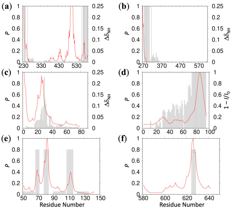
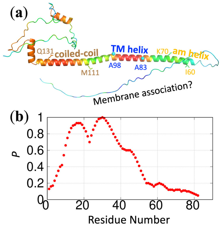
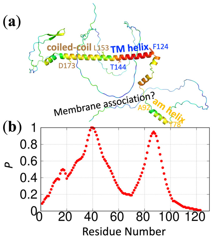
The functional processes of many proteins involve the association of their intrinsically disordered regions (IDRs) with acidic membranes. We have identified the membrane-association characteristics of IDRs using extensive molecular dynamics (MD) simulations and validated them with NMR spectroscopy. These studies have led to not only deep insight into functional mechanisms of IDRs but also to intimate knowledge regarding the sequence determinants of membrane-association propensities. Here we turned this knowledge into a web server called ReSMAP, for predicting the residue-specific membrane-association propensities from IDR sequences. The membrane-association propensities are calculated from a sequence-based partition function, trained on the MD simulation results of seven IDRs. Robustness of the prediction is demonstrated by leaving one IDR out of the training set. We anticipate there will be many applications for the ReSMAP web server, including rapid screening of IDR sequences for membrane association.
Keywords: amphipathic helix; intrinsically disordered proteins; intrinsically disordered regions; membrane binding; membrane-association propensity.
Conflict of interest statement
The authors declare no conflict of interest. The funders had no role in the design of the study; in the collection, analyses, or interpretation of data; in the writing of the manuscript; or in the decision to publish the results.
Figures

Similar articles
-
PUNCH: An Interactive Web Server for Predicting Intrinsically Disordered Regions in Protein Sequences.J Mol Biol. 2025 Aug 1;437(15):169018. doi: 10.1016/j.jmb.2025.169018. Epub 2025 Feb 21. J Mol Biol. 2025. PMID: 40133791
-
Prediction of protein-protein interactions using sequences of intrinsically disordered regions.Proteins. 2023 Jul;91(7):980-990. doi: 10.1002/prot.26486. Epub 2023 Mar 20. Proteins. 2023. PMID: 36908253
-
TransDFL: Identification of Disordered Flexible Linkers in Proteins by Transfer Learning.Genomics Proteomics Bioinformatics. 2023 Apr;21(2):359-369. doi: 10.1016/j.gpb.2022.10.004. Epub 2022 Oct 19. Genomics Proteomics Bioinformatics. 2023. PMID: 36272675 Free PMC article.
-
Towards Decoding the Sequence-Based Grammar Governing the Functions of Intrinsically Disordered Protein Regions.J Mol Biol. 2021 Jun 11;433(12):166724. doi: 10.1016/j.jmb.2020.11.023. Epub 2020 Nov 26. J Mol Biol. 2021. PMID: 33248138 Review.
-
Membrane Association of Intrinsically Disordered Proteins.Annu Rev Biophys. 2025 May;54(1):275-302. doi: 10.1146/annurev-biophys-070124-092816. Epub 2025 Feb 14. Annu Rev Biophys. 2025. PMID: 39952269 Review.
Cited by
-
Conformational ensemble-dependent lipid recognition and segregation by prenylated intrinsically disordered regions in small GTPases.Commun Biol. 2023 Nov 2;6(1):1111. doi: 10.1038/s42003-023-05487-6. Commun Biol. 2023. PMID: 37919400 Free PMC article.
-
Predicting the Sequence-Dependent Backbone Dynamics of Intrinsically Disordered Proteins.bioRxiv [Preprint]. 2024 Oct 1:2023.02.02.526886. doi: 10.1101/2023.02.02.526886. bioRxiv. 2024. Update in: Elife. 2024 Oct 30;12:RP88958. doi: 10.7554/eLife.88958. PMID: 36778236 Free PMC article. Updated. Preprint.
-
Mycobacterium tuberculosis CrgA Forms a Dimeric Structure with Its Transmembrane Domain Sandwiched between Cytoplasmic and Periplasmic β-Sheets, Enabling Multiple Interactions with Other Divisome Proteins.bioRxiv [Preprint]. 2025 Jan 16:2024.12.05.627054. doi: 10.1101/2024.12.05.627054. bioRxiv. 2025. Update in: J Am Chem Soc. 2025 Apr 02;147(13):11117-11131. doi: 10.1021/jacs.4c17168. PMID: 39677619 Free PMC article. Updated. Preprint.
-
Why Does Synergistic Activation of WASP, but Not N-WASP, by Cdc42 and PIP2 Require Cdc42 Prenylation?J Mol Biol. 2023 Apr 15;435(8):168035. doi: 10.1016/j.jmb.2023.168035. Epub 2023 Feb 28. J Mol Biol. 2023. PMID: 36863659 Free PMC article.
-
An Arg/Ala-rich helix in the N-terminal region of M. tuberculosis FtsQ is a potential membrane anchor of the Z-ring.Commun Biol. 2023 Mar 23;6(1):311. doi: 10.1038/s42003-023-04686-5. Commun Biol. 2023. PMID: 36959324 Free PMC article.
References
Grants and funding
LinkOut - more resources
Full Text Sources
