. 2022 Sep 14;96(17):e0061222.
doi: 10.1128/jvi.00612-22. Epub 2022 Aug 25.

Multiple-Site SUMOylation of FMDV 3C Protease and Its Negative Role in Viral Replication

Affiliations

Multiple-Site SUMOylation of FMDV 3C Protease and Its Negative Role in Viral Replication

Xiangju Wu et al. J Virol. .

Abstract

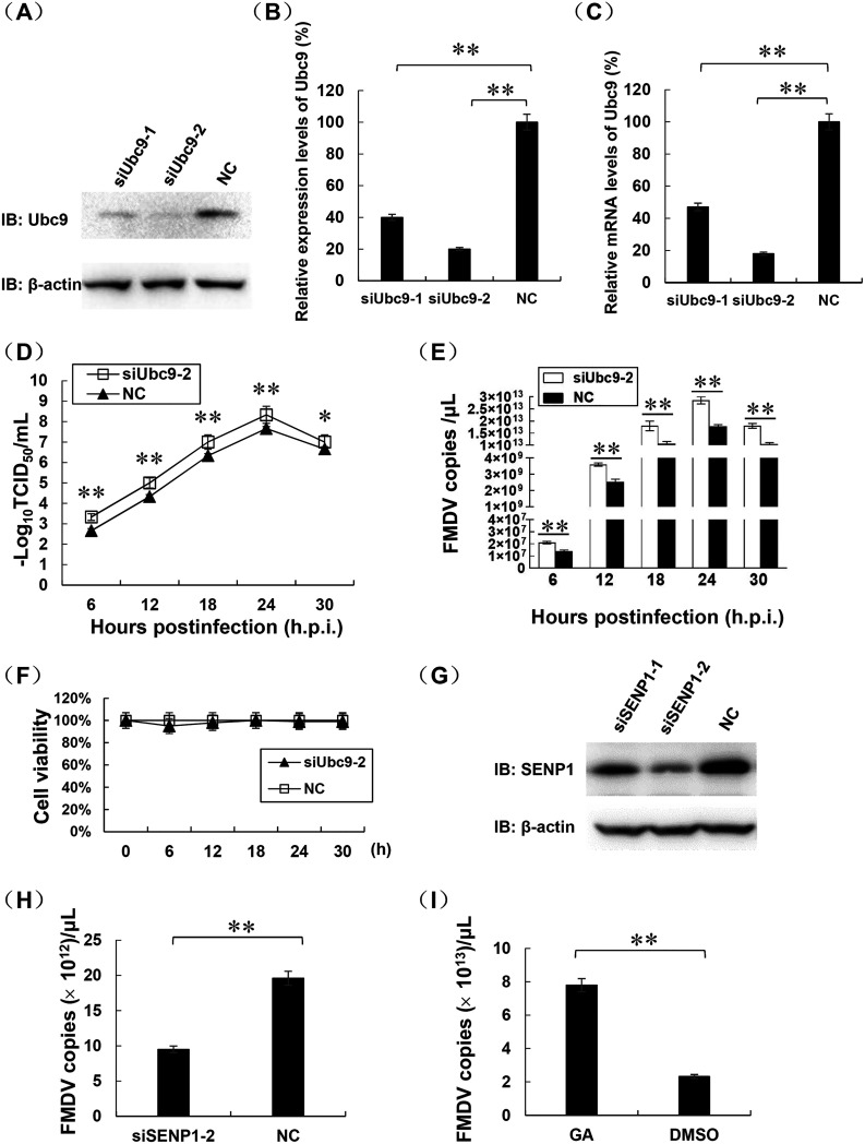
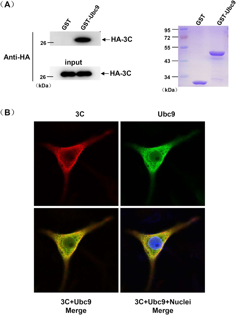
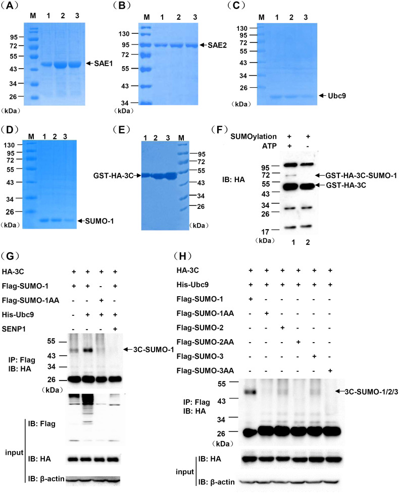
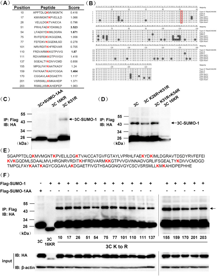
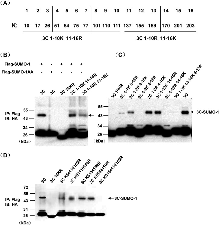
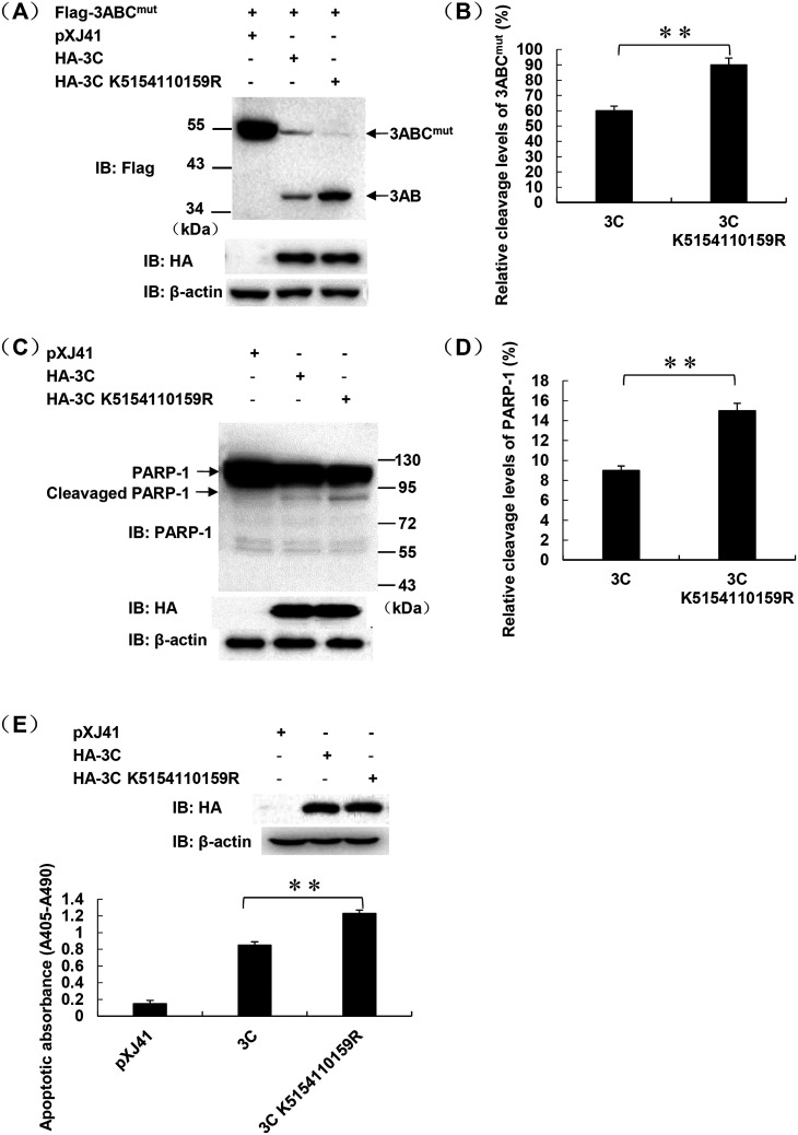
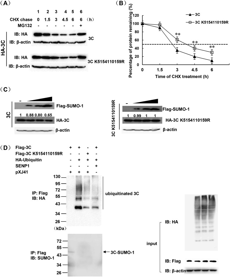
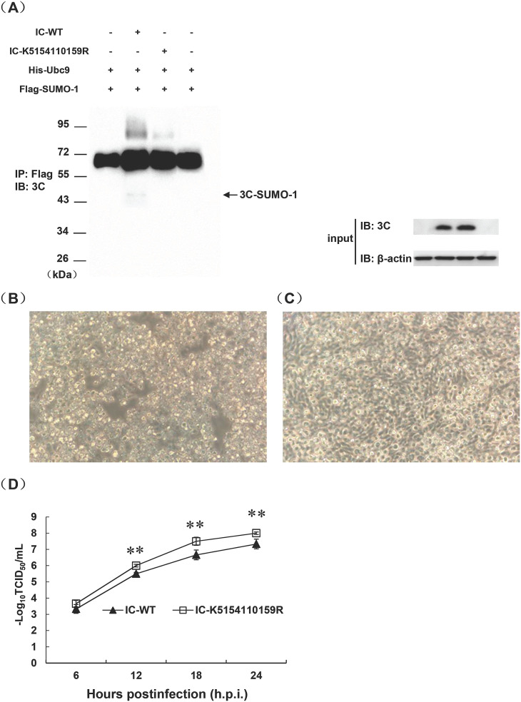
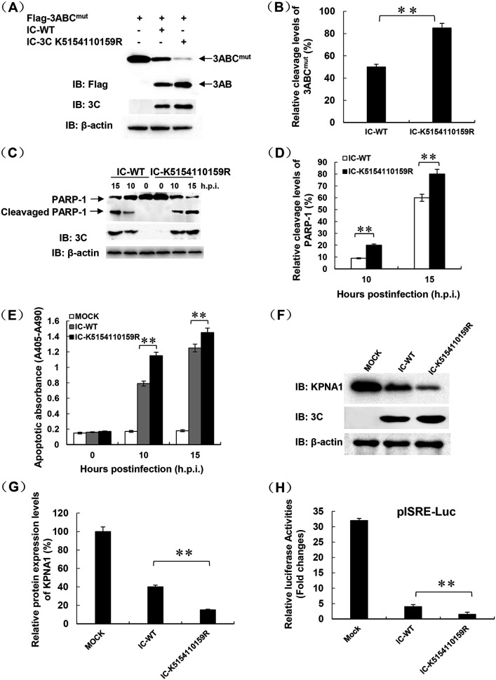
Protein SUMOylation represents an important cellular process that regulates the activities of numerous host proteins as well as of many invasive viral proteins. Foot-and-mouth disease virus (FMDV) is the first animal virus discovered. However, whether SUMOylation takes place during FMDV infection and what role it plays in FMDV pathogenesis have not been investigated. In the present study, we demonstrated that SUMOylation suppressed FMDV replication by small interfering RNA (siRNA) transfection coupled with pharmaceutical inhibition of SUMOylation, which was further confirmed by increased virus replication for SUMOylation-deficient FMDV with mutations in 3C protease, a target of SUMOylation. Moreover, we provided evidence that four lysine residues, Lys-51, -54, -110, and -159, worked together to confer the SUMOylation to the FMDV 3C protease, which may make SUMOylation of FMDV 3C more stable and improve the host's chance of suppressing the replication of FMDV. This is the first report that four lysine residues can be alternatively modified by SUMOylation. Finally, we showed that SUMOylation attenuated the cleavage ability, the inhibitory effect of the interferon signaling pathway, and the protein stability of FMDV 3C, which appeared to correlate with a decrease in FMDV replication. Taken together, the results of our experiments describe a novel cellular regulatory event that significantly restricts FMDV replication through the SUMOylation of 3C protease. IMPORTANCE FMD is a highly contagious and economically important disease in cloven-hoofed animals. SUMOylation, the covalent linkage of a small ubiquitin-like protein to a variety of substrate proteins, has emerged as an important posttranslational modification that plays multiple roles in diverse biological processes. In this study, four lysine residues of FMDV 3C were found to be alternatively modified by SUMOylation. In addition, we demonstrated that SUMOylation attenuated FMDV 3C function through multiple mechanisms, including cleavage ability, the inhibitory effect of the interferon signaling pathway, and protein stability, which, in turn, resulted in a decrease of FMDV replication. Our findings indicate that SUMOylation of FMDV 3C serves as a host cell defense against FMDV replication. Further understanding of the cellular and molecular mechanisms driving this process should offer novel insights to design an effective strategy to control the dissemination of FMDV in animals.

Keywords: 3C protease; SUMOylation; foot-and-mouth disease virus (FMDV); replication.


Conflict of interest statement

The authors declare no conflict of interest.

Figures

FIG 1
FIG 1
SUMOylation suppresses FMDV replication. (A) Western blotting of siRNA-mediated Ubc9 knockdown cells. BHK-21 cells were transfected with siRNA (siUbc9-1, siUbc9-2) or negative control siRNA (NC). Cell lysates were prepared at 24 h posttransfection and subjected to Western blotting. Endogenous Ubc9 was detected using anti-Ubc9 antibody. β-Actin was used as a protein-loading control. (B) Densitometry analysis of the digital image of endogenous Ubc9 in BHK-21 cells transfected with siRNA from three independent experiments. The band intensities of blotting with anti-Ubc9 antibody are shown as the relative protein expression levels, normalized with β-actin. (C) Quantitative RT-PCR analysis of the efficiency of siRNA targeting Ubc9. Relative transcript levels of Ubc9 are shown as relative percentage changes in comparison with the level for NC-transfected cells. (D) FMDV titers in siUbc9-2-transfected or NC-transfected cells at indicated time points. (E) Absolute quantitative RT-PCR analysis of FMDV genome copies in siUbc9-2 or NC-transfected cells. (F) Cell cytotoxicity induced by siRNA. BHK-21 cells were seeded in 96-well plates and then transfected with siUbc9-2 and NC, respectively. Twenty-four hours later, cell viability was determined at indicated time points using CellTiter 96 AQueous one-solution cell proliferation assay according to the manufacturer’s instructions. (G) Western blotting of endogenous SENP1 in siRNA-mediated knockdown cells. BHK-21 cells were transfected with siRNA (siSENP1-1, siSENP1-2) or negative control siRNA (NC). The cell lysates were prepared at 24 h posttransfection and subjected to Western blotting. Endogenous SENP1 was detected using anti-SENP1 antibody. β-Actin was used as a protein-loading control. (H) Absolute quantitative RT-PCR analysis of FMDV genome copies in siSENP1-2 or NC-transfected cells at 24 hpi. (I) Absolute quantitative RT-PCR analysis of FMDV genome copies in glutaraldehyde (GA)- or dimethyl sulfoxide (DMSO)-treated BHK-21 cells at 24 hpi. Data represent the means ± the standard deviations (error bars) of three independent experiments. *, P < 0.05; **, P < 0.01.
FIG 2
FIG 2
FMDV 3C and Ubc9 interact in vitro and colocalize in the nucleus and cytoplasm. (A) GST pulldown analysis of FMDV 3C and Ubc9 interaction. Western blotting of HA-3C proteins pulled down by GST or GST-Ubc9 using anti-HA antibody (top). Input represents HA-3C protein expressed in HEK-293T cells (bottom). Coomassie blue staining shows GST and GST-Ubc9 proteins used for each binding reaction (right). (B) Confocal microscopy for colocalization of FMDV 3C and Ubc9. BHK-21 cells were cotransfected with pXJ41-HA-3C and pXJ41-His-Ubc9. At 18 h posttransfection, cells were fixed, and confocal microscopy was conducted as described in Materials and Methods. HA-3C and His-Ubc9 proteins were stained red and green, respectively. Nuclei were stained blue.
FIG 3
FIG 3
FMDV 3C is mainly SUMOylated by SUMO-1. (A to E) Coomassie blue staining shows each component of in vitro SUMOylation assay purified using Ni-nitrilotriacetic acid (NTA) or glutathione-Sepharose 4B agarose, including His-SAE1 (A), His-SAE2 (B), His-Ubc9 (C), His-SUMO-1 (D), and GST-HA-3C (E); lanes 1 to 3 represent different eluents. (F) Western blotting for in vitro SUMOylation of GST-HA-3C protein. SUMO-modified and unmodified GST-HA-3C proteins were indicated with arrows. “SUMOylation” represents samples incubated with all components in the SUMOylation reaction except ATP. (G) In vivo SUMOylation of FMDV 3C protein by SUMO-1 in HEK-293T cells. The SUMOylation band of 3C protein is marked with an arrow. (H) In vivo SUMOylation of 3C protein by SUMO-1, SUMO-2, or SUMO-3 in HEK-293T cells. The SUMO-modified 3C protein is indicated by an arrow. The data presented here are results from one experiment of three independent Western blotting experiments.
FIG 4
FIG 4
Coimmunoprecipitation (co-IP) and Western blotting of FMDV 3C SUMOylation in FMDV-infected cells. BHK-21 cells were cotransfected with pXJ41-His-Ubc9 and pXJ41-Flag-SUMO-1. Twenty-four hours later, cells were infected with FMDV and lysed at indicated postinfection time points. (A) Lysates were immunoprecipitated and probed with rabbit anti-3C polyclonal antibody. Arrow indicates the SUMOylated FMDV 3C, and arrowhead indicates FMDV 3C. (B) Lysates were immunoprecipitated with anti-Flag M2 beads followed by immunoblot using rabbit anti-3C polyclonal antibody. Arrow indicates SUMOylated FMDV 3C. (C) Western blotting of the lysates using rabbit anti-3C polyclonal antibody. Arrow indicates the SUMOylated FMDV 3C, and arrowhead indicates FMDV 3C. (D) Western blotting of the lysates using anti-Flag antibody. Arrowhead indicates Flag-SUMO-1.
FIG 5
FIG 5
A single Lys of 3C is insufficient to confer SUMO conjugation. (A) Bioinformatics prediction of potential SUMOylation sites in FMDV 3C protein using GPS-SUMO 2.0 Online Service. (B) Comparison of the 3C amino acid sequence of strains from all of the 7 serotypes of FMDV. (C, D, and F) SUMOylation of 3C or its mutants in HEK-293T cells. The lysates of HEK-293T cells were coimmunoprecipitated with anti-Flag M2 beads and then subjected to Western blotting with anti-HA antibody. Input, whole-cell lysates. SUMOylation band is marked with an arrow. (E) Amino acid sequence of FMDV 3C. Lys residues mutated to Arg for SUMOylation analysis are shown in bold and red.
FIG 6
FIG 6
Lys-51, -54, -110, and -159 are FMDV 3C SUMOylation sites. (A) Positions (bottom numbers) of Lys residues in 3C and the numbering (top numbers) of each Lys. Horizontal lines divide 3C into two parts, and vertical lines divide 3C into four parts. 3C mutants were constructed by replacing Lys with Arg according to this schematic presentation of Lys residues in 3C. (B to D) SUMOylation of 3C mutants in HEK-293T cells. SUMOylation band is marked with an arrow.
FIG 7
FIG 7
The cleavage ability of FMDV 3C is downregulated by SUMOylation. (A) SUMOylation downregulates the polyprotein precursor cleavage efficiency of FMDV 3C. HEK-293T cells were cotransfected with Flag-3ABCmut and wild-type 3C (WT-3C) or SUMOylation-deficient 3C quadruple mutant (3C K5154110159R). pXJ41 was used as negative control. At 24 h posttransfection, cells were lysed and subjected to Western blotting using antibody against Flag, HA, or β-actin, respectively. The intact 3ABCmut or its cleavaged product was marked by arrows. (B) The optical density ratios of 3AB over total 3ABCmut proteins in panel A are shown in graphs. (C) SUMOylation downregulates the PARP-1 cleavage induced by FMDV 3C. HEK-293T cells were transfected with WT-3C or SUMOylation-deficient 3C quadruple mutant. pXJ41 was used as a negative control. At 24 h posttransfection, cells were lysed and subjected to Western blotting using antibody against endogenous PARP-1, HA, or β-actin, respectively. (D) The optical density ratios of cleavaged PARP-1 over total PARP-1 proteins in panel C are shown in graphs. (E) Apoptosis of HEK-293T cells transfected with WT-3C or SUMOylation-deficient 3C quadruple mutant was quantified by cell death detection ELISA. Expression of 3C or β-actin in HEK-293T cells was detected by Western blotting using antibody against HA or β-actin. Data represent the means ± the standard deviations (error bars) of three independent experiments. **, P < 0.01.
FIG 8
FIG 8
The inhibitory effect of interferon signaling pathway induced by FMDV 3C is attenuated by SUMOylation at the protein level. (A) SUMOylation attenuates KPNA1 degradation induced by 3C. HeLa cells were cotransfected with Flag-KPNA1 and WT-3C or SUMOylation-deficient 3C quadruple mutant. pXJ41 was used as a negative control. At 24 h posttransfection, cells were lysed and subjected to Western blotting using antibody against Flag, HA, or β-actin, respectively. (B) The optical density ratios of KPNA1/β-actin in panel A are shown in graphs. (C) SUMOylation attenuates the inhibition of mRNA levels of ISGs induced by 3C. HeLa cells were transfected with pXJ41, pXJ41-HA-3C, or pXJ41-HA-3C quadruple mutant and treated with 1,000 U/mL of IFN-β at 24 h posttransfection. Five hours later, the transcript levels of ISGs were analyzed by real-time RT-PCR. pXJ41 was used as a negative control. Relative transcript levels are shown as relative fold changes in comparison with the level for the mock-treated control of untransfected cells. Expression of 3C, 3C quadruple mutant, or β-actin in HeLa cells was detected by Western blotting using antibody against HA or β-actin. (D) SUMOylation attenuates the inhibition of ISRE promoter activity induced by 3C. HeLa cells were cotransfected with 0.5 μg of pXJ41, pXJ41-HA-3C, or pXJ41-HA-3C quadruple mutant and 0.5 μg of pISRE-Luc along with 0.05 μg of pRL-TK. A luciferase reporter gene assay was conducted as described in Materials and Methods. Expression of 3C, 3C quadruple mutant, or β-actin in HeLa cells was detected by Western blotting using antibody against HA or β-actin. All assays were repeated three times, with each experiment performed in triplicate. Data represent the means ± the standard deviations (error bars) of three independent experiments. *, P < 0.05; **, P < 0.01.
FIG 9
FIG 9
SUMOylation promotes 3C protein degradation. (A) Western blotting of WT-3C or 3C quadruple mutant (3C K5154110159R) expression in HEK-293T cells treated with 100 μg/mL cycloheximide (CHX) for the indicated times in the presence or absence of 10 mM MG132. (B) The optical density ratios of 3C/β-actin in panel A are shown in graphs. The plot shows quantification of the half-life of WT-3C or 3C quadruple mutant (3C K5154110159R) protein from three independent experiments by nonlinear regression analysis. Data represent the means ± the standard deviations (error bars) of three experiments. **, P < 0.01. (C) Western blotting of WT-3C or 3C quadruple mutant (3C K5154110159R) stability in HEK-293T cells cotransfected with increasing amounts (wedge) of pXJ41-Flag-SUMO-1 plasmid or pXJ41 empty vector (−). The amount of 3C in pXJ41-Flag-SUMO-1-cotransfected cells relative to that in pXJ41-cotransfected cells (lane 1) is indicated at the top of each band. (D) Immunoblot analysis with anti-HA (left, top) or anti-SUMO-1 (left, bottom) for proteins coimmunoprecipitated with anti-Flag M2 beads from lysates of HEK-293T cells. Cells were cotransfected with plasmids as indicated. Input, whole-cell lysates.
FIG 10
FIG 10
SUMOylation of 3C attenuates FMDV replication. (A) BHK-21 cells were transfected with the indicated plasmids for 24 h and then infected with FMDV IC-WT or FMDV quadruple mutant IC-K5154110159R at an MOI of 0.01. Ten hours later, cells were lysed and immunoprecipitated with anti-Flag M2 beads followed by immunoblot using rabbit anti-3C polyclonal antibody. Arrow indicates sumoylated 3C. Input, whole-cell lysates. (B) CPE induced by FMDV quadruple mutant IC-K5154110159R at 12 hpi in BHK-21 cells (magnification, ×200). (C) CPE induced by FMDV IC-WT at 12 hpi in BHK-21 cells (magnification, ×200). (D) Growth kinetics of FMDV IC-WT and FMDV quadruple mutant IC-K5154110159R. Data represent the means ± the standard deviations (error bars) of three independent experiments. **, P < 0.01.
FIG 11
FIG 11
The cleavage ability and the inhibitory effect of the interferon signaling pathway of FMDV 3C are attenuated by SUMOylation at the viral level. (A) PK-15 cells transfected with Flag-3ABCmut were infected with FMDV IC-WT or FMDV quadruple mutant IC-K5154110159R at an MOI of 0.01 and harvested at 24 hpi for Western blotting using antibody against Flag, 3C, or β-actin. (B) The optical density ratios of 3AB over total 3ABCmut proteins in panel A are shown in graphs. (C) PK-15 cells were infected with FMDV IC-WT or FMDV quadruple mutant IC-K5154110159R at an MOI of 0.01 and harvested at 0, 10, and 15 hpi for Western blotting using antibody against PARP-1, 3C, or β-actin. (D) The optical density ratios of cleavaged PARP-1 over total PARP-1 proteins in panel C are shown in graphs. (E) Apoptosis was quantified by cell death detection ELISA. (F) PK-15 cells were infected with FMDV IC-WT or FMDV quadruple mutant IC-K5154110159R at an MOI of 0.01 and harvested at 24 hpi for Western blotting using antibody against KPNA1, 3C, or β-actin, respectively. (G) The optical density ratios of KPNA1/β-actin in panel F are shown in graphs. (H) PK-15 cells were cotransfected with 0.5 μg of pISRE-Luc and 0.05 μg of pRL-TK. Twelve hours later, cells were infected with FMDV IC-WT or FMDV quadruple mutant IC-K5154110159R at an MOI of 0.01. A luciferase reporter gene assay was conducted as described in Materials and Methods. All assays were repeated three times, with each experiment performed in triplicate. Data represent the means ± the standard deviations (error bars) of three independent experiments. **, P < 0.01.
FIG 12
FIG 12
The protein stability of FMDV 3C is attenuated by SUMOylation at the viral level. (A) PK-15 cells were infected with FMDV IC-WT or FMDV quadruple mutant IC-K5154110159R at an MOI of 0.01, and 24 h later, cells were treated with 150 μg/mL CHX for the indicated times and subjected to Western blotting using antibody against 3C or β-actin. (B) The optical density ratios of 3C/β-actin in panel A are shown in graphs. The plot shows quantification of the 3C protein half-life from three independent experiments by nonlinear regression analysis. (C) PK-15 cells were infected with FMDV IC-WT or FMDV quadruple mutant IC-K5154110159R at an MOI of 0.01. Ten hours later, lysates were immunoprecipitated with rabbit anti-3C polyclonal antibody followed by immunoblot using mouse anti-ubiquitin monoclonal antibody. Input, whole-cell lysates. Data represent the means ± the standard deviations (error bars) of three independent experiments. **, P < 0.01.

Similar articles

Cited by

References

    1. Fry EE, Stuart DI, Rowlands DJ. 2005. The structure of foot-and-mouth disease virus. Curr Top Microbiol Immunol 288:71–101. 10.1007/3-540-27109-0_4. - DOI - PubMed
    1. Grubman M, Baxt B. 2004. Foot-and-mouth disease. Clin Microbiol Rev 17:465–493. 10.1128/CMR.17.2.465-493.2004. - DOI - PMC - PubMed
    1. Belsham GJ. 2005. Translation and replication of FMDV RNA. Curr Top Microbiol Immunol 288:43–70. 10.1007/3-540-27109-0_3. - DOI - PubMed
    1. Mason PW, Grubman MJ, Baxt B. 2003. Molecular basis of pathogenesis of FMDV. Virus Res 91:9–32. 10.1016/s0168-1702(02)00257-5. - DOI - PubMed
    1. Birtley JR, Knox SR, Jaulent AM, Brick P, Leatherbarrow RJ, Curry S. 2005. Crystal structure of foot-and-mouth disease virus 3C protease: new insights into catalytic mechanism and cleavage specificity. J Biol Chem 280:11520–11527. 10.1074/jbc.M413254200. - DOI - PubMed

LinkOut - more resources