. 2022 Aug 11;5(4):67.
doi: 10.3390/mps5040067.

Laser Microdissection-Mediated Isolation of Butterfly Wing Tissue for Spatial Transcriptomics

Affiliations

Laser Microdissection-Mediated Isolation of Butterfly Wing Tissue for Spatial Transcriptomics

Tirtha Das Banerjee et al. Methods Protoc. .

Abstract

The assignment of specific patterns of gene expression to specific cells in a complex tissue facilitates the connection between genotype and phenotype. Single-cell sequencing of whole tissues produces single-cell transcript resolution but lacks the spatial information of the derivation of each cell, whereas techniques such as multiplex FISH localize transcripts to specific cells in a tissue but require a priori information of the target transcripts to examine. Laser dissection of tissues followed by transcriptome analysis is an efficient and cost-effective technique that provides both unbiased gene expression discovery together with spatial information. Here, we detail a laser dissection protocol for total RNA extraction from butterfly larval and pupal wing tissues, without the need of paraffin embedding or the use of a microtome, that could be useful to researchers interested in the transcriptome of specific areas of the wing during development. This protocol can bypass difficulties in extracting high quality RNA from thick fixed tissues for sequencing applications.

Keywords: Bicyclus anynana; RNAseq; eyespots; laser microdissection; spatial transcriptomics; wing sectors.


Conflict of interest statement

The authors declare no conflict of interest.

Figures

Figure 1
Figure 1
Steps involved in the microdissection of butterfly larval and pupal wing tissue. Step 1 involves the transfer of the wing tissue onto the PEN membrane slides and fixation using ethanol and acetone. Step 2 involves staining the wings with Histogene solution and performing washes with ethanol. Step 3 involves laser microdissection using a Zeiss PALM microbeam. Step 4 involves isolation of the microsections from the dissected microtissues using a pair of fine tweezers and transfer of microsections into molecular grade water. Afterwards, the tissues can be stored at −80 °C. Step 5 involves extraction of RNA from the microdissected tissues and testing the integrity of the RNA.
Figure 2
Figure 2
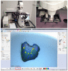
Zeiss PALM microbeam-laser microdissection microscope and PALMRobo 4.8 Pro settings for laser microdissection of Bicyclus anynana larval and pupal wings. (A) Zeiss PALM microbeam-laser microdissection microscope. (B) A closeup of the mounted slide on the microscope. (C) Settings used for laser microdissection of the larval and pupal wing tissues. Magenta arrow: slide holder control; yellow arrow: collection cap control; orange arrow: laser energy; red arrow: laser focus; green arrow: number of laser cutting cycles; blue arrow: intensity for the backlight; and red box: tools for selecting the sections for microdissection.
Figure 3
Figure 3
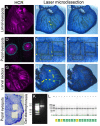
Laser microdissection of larval and pupal wings. (A) Larval wings showing the expression of the gene spalt in the center of the eyespots (visualized with a hybridization chain reaction). (B,C) Panel A is used as a reference to localize the position of the eyespot center in a wing at a similar developmental stage (B,C) and to perform the microdissections. (D) Pupal wing, expressing the genes spalt (magenta) and optix (cyan) in the rings of the eyespots (hybridization chain reaction) is used as a reference to localize the position of the eyespots in a wing at a similar developmental stage (E,F) to perform microdissections. (G) Larval wing showing expression of the gene spalt in the different sectors of the wings (hybridization chain reaction) is used as a reference to localize the positions of the wing sectors along with the venation pattern on a wing at a similar developmental stage (H,I) to perform microdissections for sector-specific transcriptomes. (J) A pupal wing with the eyespot regions removed with a pair of fine tweezers. (K) An agarose gel electrophoresis on the RNA extracted from laser-dissected wing tissue with a clear band of RNA. (L) RIN value of different RNA samples from the larval eyespot and control regions from wet season (WS) and dry season (DS) forms of B. anynana butterflies. A0: ladder; A1, C1, E1, G1: WS control; A2, C2, E2, G2: DS control; B1, D1, F1, H1: WS eyespot; B2, D2, F2, H2: DS eyespot. Green line: lower marker. The samples were sent for RNA-seq and the gel image was generated from Azenta, Singapore.
Figure 4
Figure 4
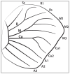
Venation nomenclature of Bicyclus anynana larval hindwing used for dissection and isolation of sector-specific transcriptomes using LCM.

Similar articles

References

    1. Chen K.H., Boettiger A.N., Moffitt J.R., Wang S., Zhuang X. Spatially Resolved, Highly Multiplexed RNA Profiling in Single Cells. Science. 2015;348:1360–1363. doi: 10.1126/science.aaa6090. - DOI - PMC - PubMed
    1. Lubeck E., Coskun A.F., Zhiyentayev T., Ahmad M., Cai L. Single-Cell in Situ RNA Profiling by Sequential Hybridization. Nat. Methods. 2014;11:360–361. doi: 10.1038/nmeth.2892. - DOI - PMC - PubMed
    1. Eng C.H.L., Lawson M., Zhu Q., Dries R., Koulena N., Takei Y., Yun J., Cronin C., Karp C., Yuan G.C., et al. Transcriptome-Scale Super-Resolved Imaging in Tissues by RNA SeqFISH+ Nature. 2019;568:235–239. doi: 10.1038/s41586-019-1049-y. - DOI - PMC - PubMed
    1. Goh J.J.L., Chou N., Seow W.Y., Ha N., Cheng C.P.P., Chang Y.C., Zhao Z.W., Chen K.H. Highly Specific Multiplexed RNA Imaging in Tissues with Split-FISH. Nat. Methods. 2020;17:689–693. doi: 10.1038/s41592-020-0858-0. - DOI - PubMed
    1. Codeluppi S., Borm L.E., Zeisel A., La Manno G., van Lunteren J.A., Svensson C.I., Linnarsson S. Spatial Organization of the Somatosensory Cortex Revealed by OsmFISH. Nat. Methods. 2018;15:932–935. doi: 10.1038/s41592-018-0175-z. - DOI - PubMed

LinkOut - more resources