. 2022 Sep 20;204(9):e0020822.
doi: 10.1128/jb.00208-22. Epub 2022 Aug 25.

Overlapping and Distinct Functions of the Paralogous PagR Regulators of Bacillus anthracis

Affiliations

Overlapping and Distinct Functions of the Paralogous PagR Regulators of Bacillus anthracis

Ileana D Corsi et al. J Bacteriol. .

Abstract

The Bacillus anthracis pagA gene, encoding the protective antigen component of anthrax toxin, is part of a bicistronic operon on pXO1 that codes for its own repressor, PagR1. In addition to the pagAR1 operon, PagR1 regulates sap and eag, two chromosome genes encoding components of the surface layer, a mounting structure for surface proteins involved in virulence. Genomic studies have revealed a PagR1 paralog, PagR2, encoded by a gene on pXO2. The amino acid sequences of the paralogues are 71% identical and show similarity to the ArsR family of transcription regulators. We determined that the expression of either rPagR1 or rPagR2 in a ΔpagR1 pXO1+/pXO2- (PagR1-PagR2) background repressed the expression of pagA, sap, eag, and a newly discovered target, atxA, encoding virulence activator AtxA. Despite the redundancy in PagR1 and PagR2 function, we determined that purified rPagR1 bound DNA corresponding to the control regions of all four target genes and existed as a dimer in cell lysates, whereas rPagR2 exhibited weak binding to the DNA of the pagA and atxA promoters, did not bind sap or eag promoter DNA, and did not appear as a dimer in cell lysates. A single amino acid change in PagR2, S81Y, designed to match the native Y81 of PagR1, allowed for DNA-binding to the sap and eag promoters. Moreover, the S81Y mutation allowed for the detection of PagR2 homomultimers in coaffinity purification experiments. Our results expand our knowledge of the roles of the paralogues in B. anthracis gene expression and provide a potential mechanistic basis for differences in the functions of these repressors. IMPORTANCE The protective antigen component of the anthrax toxin is essential for the delivery of the enzymatic components of the toxin into host target cells. The toxin genes and other virulence genes of B. anthracis are regulated by multiple trans-acting regulators that respond to a variety of host-related signals. PagR1, one such trans-acting regulator, connects the regulation of plasmid-encoded and chromosome-encoded virulence genes by controlling both protective antigen and surface layer protein expression. Whether PagR2, a paralog of PagR1, also functions as a trans-acting regulator was unknown. This work advances our knowledge of the complex model of virulence regulation in B. anthracis and furthers our understanding of the intriguing evolution of this pathogen.

Keywords: ArsR family; Bacillus anthracis; anthrax toxin; paralogous proteins; regulatory paralogs; transcriptional regulation.


Conflict of interest statement

The authors declare no conflict of interest.

Figures

FIG 1
FIG 1
Comparison of PagR1 and PagR2 amino acid sequence. (A) Amino acid sequence alignment of PagR1 and PagR2. Both proteins are 99 amino acids long. Amino acid residues shaded in black boxes indicate conserved identical residues between the two proteins, while those shaded in gray boxes indicate similar residues belonging to the same group. Unshaded amino acids are completely different between the two proteins and are therefore not conserved. Amino acids predicted to participate in DNA binding, according to the published PagR1 crystal structure (26), and in the conservation of ArsR-family protein residues are labeled in red squares. Regions of secondary structure that were elucidated from the PagR1 crystal structure are shown above the aligned sequences. The sequence alignment was generated using the T-Coffee and Boxshade webservers. (B) Comparison of PagR1 and PagR2 secondary structure, as modeled using PyMOL 2.5 software. The predicted PagR2 secondary structure closely matches the published PagR1 crystal structure (26). Predicted secondary structure features are labeled as follows: α-helix 1 (H1: dark blue), α-helix 2 (H2: light blue), α-helix 3 (H3: green), α-helix 4 (H4: yellow), two β-sheets forming a β-sheet hairpin (β1 and β2: orange), and α-helix 5 (H5: pink).
FIG 2
FIG 2
Effect of PagR1 and PagR2 on virulence gene expression. Total RNA isolated from cell lysates was used to probe for (A) pagA, (B) atxA, (C) sap, and (D) eag expression using TaqMan-based qRT-PCR assays. The mRNA levels in each strain were normalized to levels of the gyrB reference mRNA. Data are presented as averages of three biological replicates with the standard deviations. An analysis of variance (ANOVA) followed by Tukey’s multiple-comparison test was used to determine statistical significance. Asterisks directly above the bars indicate a statistically significant difference between that bar and the “Parent” bar. Additional comparisons are indicated with brackets, with the respective significance shown by asterisks above the bracket. * indicates P-value < 0.05, ** indicates < 0.01, *** indicates < 0.001, and **** indicates < 0.0001.
FIG 3
FIG 3
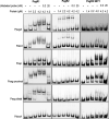
Specific promoter binding by PagR1 and PagR2 proteins. Purified recombinant PagR1, PagR2, and PagR2 S81Y proteins were incubated at the indicated, increasing concentrations with 0.2 nM biotin-labeled probes. The probes were made via the PCR amplification of the promoters of pagA, atxA, sap, and eag (both the distal and the proximal promoters). The promoter of dnaA was used as a negative-control for the no binding interaction. Unlabeled probes were added to the indicated binding reactions at a final concentration of either 0.2 or 20 nM. The blots shown are representative images of three replicates.
FIG 4
FIG 4
Comparison of PagR1 and PagR2 Kds. Purified recombinant PagR1 and PagR2 were incubated at increasing concentrations with 0.2 nM biotin-labeled probes. The probe binding of (A) Psap and (B) Peag proximal were calculated as a fraction of bound promoter DNA over the total promoter DNA. Increasing binding with increasing protein concentration was plotted, and Kds were calculated using the Hill equation. Kds are reported with 95% confidence intervals.
FIG 5
FIG 5
Homomultimerization of recombinant PagR proteins. Multimerization of recombinant proteins was explored using coaffinity purification. Cell lysates from ΔpagR1 strains expressing either His- or FLAG-tagged PagR proteins were coincubated as indicated and subjected to affinity purification using Ni-NTA resin. Coincubation with GFP-FLAG was performed as a negative-control. Proteins present prior to (Input) and after (Eluate) purification were detected using Western blotting with α-His and α-FLAG antibodies. Detection of FLAG-tagged proteins post Ni-NTA purification was indicative of interaction between the His- and FLAG-tagged coincubated proteins. (A) His- and FLAG-tagged PagR1. (B) His- and FLAG-tagged PagR2. (C) His- and FLAG-tagged PagR2 S81Y mutant.
FIG 6
FIG 6
Heteromultimerization of recombinant PagR proteins. Cell lysates from ΔpagR1 strains expressing either PagR1-FLAG or PagR2-His were coincubated as indicated and subjected to affinity purification using Ni-NTA resin. Proteins present prior to (Input) and after (Eluate) purification were detected using Western blotting with α-His and α-FLAG antibodies. (A) PagR2-His and PagR1-FLAG. (B) PagR2 S81Y-His mutant and PagR1-FLAG.
FIG 7
FIG 7
Model of virulence gene regulation by trans-acting paralogs. The pagR1 and pagR2 loci are shown in red bolded letters on the pXO1 and pXO2 plasmids, respectively. The master virulence regulator of B. anthracis, AtxA, directly activates the expression of the pagAR1 locus. PagR1 strongly represses pagA, atxA, and sap. PagR1 also represses one of the two eag promoters, allowing for preferential transcription initiation by an unknown pXO1 factor at the alternative promoter. AtxA and its pXO2-encoded paralogs, AcpA and AcpB, positively control the pagR2 locus. Purified PagR2 weakly bound the pagA and atxA promoters, as shown with solid arrows. The PagR-mediated control of atxA suggests autogenous regulation of the pagR loci. PagR2 appears to indirectly affect sap and eag expression, as shown with dashed arrows. PagR1 is shown as a dimer, while PagR2 is shown as a monomer. The surface-exposed PagR1 Y81 may mediate protein-protein interactions that facilitate PagR1 dimerization and influence DNA-binding activity. PagR2 S81 residue appears to be buried in the secondary structure. The mutation of PagR2 S81 to Y facilitates protein-protein interactions and the DNA-binding activity of PagR2.

Similar articles

Cited by

References

    1. Baker CR, Hanson-Smith V, Johnson AD. 2013. Following gene duplication, paralog interference constrains transcriptional circuit evolution. Science 342:104–108. 10.1126/science.1240810. - DOI - PMC - PubMed
    1. Price MN, Dehal PS, Arkin AP. 2008. Horizontal gene transfer and the evolution of transcriptional regulation in Escherichia coli. Genome Biol 9:R4. 10.1186/gb-2008-9-1-r4. - DOI - PMC - PubMed
    1. Fortuna A, Bähre H, Visca P, Rampioni G, Leoni L. 2021. The two Pseudomonas aeruginosa DksA stringent response proteins are largely interchangeable at the whole transcriptome level and in the control of virulence-related traits. Environ Microbiol 23:5487–5504. 10.1111/1462-2920.15693. - DOI - PubMed
    1. Chanda PK, Bandhu A, Jana B, Mondal R, Ganguly T, Sau K, Lee CY, Chakrabarti G, Sau S. 2010. Characterization of an unusual cold shock protein from Staphylococcus aureus. J Basic Microbiol 50:519–526. 10.1002/jobm.200900264. - DOI - PMC - PubMed
    1. Gallegos-Monterrosa R, Christensen MN, Barchewitz T, Koppenhöfer S, Priyadarshini B, Bálint B, Maróti G, Kempen PJ, Dragoš A, Kovács ÁT. 2021. Impact of Rap-Phr system abundance on adaptation of Bacillus subtilis. Commun Biol 4. 10.1038/s42003-021-01983-9. - DOI - PMC - PubMed

Publication types

MeSH terms

LinkOut - more resources