. 2022 Oct 27;34(11):4583-4599.
doi: 10.1093/plcell/koac266.

The localization of PHRAGMOPLAST ORIENTING KINESIN1 at the division site depends on the microtubule-binding proteins TANGLED1 and AUXIN-INDUCED IN ROOT CULTURES9 in Arabidopsis

Affiliations

The localization of PHRAGMOPLAST ORIENTING KINESIN1 at the division site depends on the microtubule-binding proteins TANGLED1 and AUXIN-INDUCED IN ROOT CULTURES9 in Arabidopsis

Alison M Mills et al. Plant Cell. .

Abstract

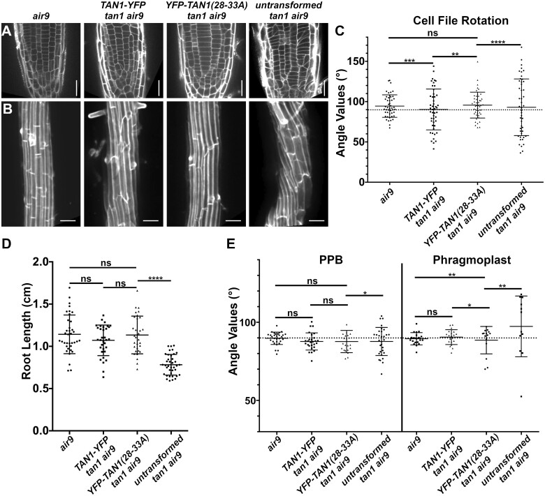
Proper plant growth and development require spatial coordination of cell divisions. Two unrelated microtubule-binding proteins, TANGLED1 (TAN1) and AUXIN-INDUCED IN ROOT CULTURES9 (AIR9), are together required for normal growth and division plane orientation in Arabidopsis (Arabidopsis thaliana). The tan1 air9 double mutant has synthetic growth and division plane orientation defects, while single mutants lack obvious defects. Here we show that the division site-localized protein, PHRAGMOPLAST ORIENTING KINESIN1 (POK1), was aberrantly lost from the division site during metaphase and telophase in the tan1 air9 mutant. Since TAN1 and POK1 interact via the first 132 amino acids of TAN1 (TAN11-132), we assessed the localization and function of TAN11-132 in the tan1 air9 double mutant. TAN11-132 rescued tan1 air9 mutant phenotypes and localized to the division site during telophase. However, replacing six amino-acid residues within TAN11-132, which disrupted the POK1-TAN1 interaction in the yeast-two-hybrid system, caused loss of both rescue and division site localization of TAN11-132 in the tan1 air9 mutant. Full-length TAN1 with the same alanine substitutions had defects in phragmoplast guidance and reduced TAN1 and POK1 localization at the division site but rescued most tan1 air9 mutant phenotypes. Together, these data suggest that TAN1 and AIR9 are required for POK1 localization, and yet unknown proteins may stabilize TAN1-POK1 interactions.


Figures

Figure 1
Figure 1
TAN1 and AIR9 together promote POK1 maintenance at the division site. YFP–POK1 localization in Col-0 WT, tan1 single mutant, air9 single mutant, and tan1 air9 double mutant plants expressing UBQ10:mScarlet-MAP4 to mark microtubules and pPOK1:YFPPOK1. Scale bars = 10 µm. A–C, YFP–POK1 localization in Col-0 WT plants. A, YFP–POK1 localization during preprophase/prophase. B, YFP–POK1 was maintained at the division site in metaphase and anaphase cells in Col-0 plants. C, YFP–POK1 remains clearly visible at the division site in Col-0 telophase cells. D–F, YFP–POK1 localization in tan1 air9 double mutant plants. D, YFP–POK1 localization during preprophase/prophase. E, YFP–POK1 was lost from the division site upon entry into metaphase. F, In tan1 air9 telophase cells YFP–POK1 was absent from the division site and accumulated in the phragmoplast midline. G–I, YFP–POK1 localization in tan1 single mutant plants. G, YFP–POK1 localization in the tan1 single mutant during preprophase/prophase. H, YFP–POK1 was maintained at the division site in metaphase and anaphase cells in tan1 plants. I, YFP–POK1 remains clearly visible at the division site in tan1 telophase cells. J–L, YFP–POK1 localization in air9 single mutant plants. J, YFP–POK1 localization during preprophase/prophase. K, YFP–POK1 was maintained at the division site in metaphase and anaphase cells in air9 plants. L, YFP–POK1 remains clearly visible at the division site in air9 telophase. M, YFP–POK1 subcellular localization in WT, tan1 air9, tan1, and air9 plants in preprophase/prophase, metaphase, and telophase cells. N > 14 plants for each genotype. Statistically significant differences in localization compared to WT plants were determined using Fisher’s exact test with Bonferroni correction for four sample types. Asterisks indicate significant differences. YFP–POK1 colocalized with the PPB in 71% of WT cells (50/70), 50% of tan1 air9 cells (27/54), 64% of tan1 cells (54/85), and 64% of air9 cells (46/72). YFP–POK1 colocalization with the PPB was not significantly different in tan1 air9, tan1, and air9 plants compared to in Wt plants. YFP–POK1 was maintained at the division site in all metaphase cells of WT (13/13 cells), tan1 (17/17 cells), and air9 (24/24 cells) plants. In tan1 air9 double mutant plants, YFP–POK1 was not maintained at the division site during metaphase (0/21 cells, P < 0.00001). During telophase, YFP–POK1 accumulated at the division site only in 87% of WT cells (27/31). In 13% of WT telophase cells, YFP–POK1 accumulated at the division site and in the phragmoplast midline (4/31 cells). YFP–POK1 localization in telophase cells of air9 single mutant plants was not significantly different from that in WT plants, with 90% of cells (36/40) accumulating YFP–POK1 at the division site only and 10% (4/40) accumulating YFP–POK1 at the division site and in the phragmoplast midline. In tan1 single mutant plants, YFP–POK1 was present at the division site in all telophase cells but accumulated more frequently at the division site and phragmoplast midline compared to in WT plants (44%; 12/27 cells; P = 0.0094). In tan1 air9 double mutant plants, YFP–POK1 never accumulated at the division site of telophase cells and instead accumulated solely in the phragmoplast midline (34/44 cells, P < 0.00001).
Figure 2
Figure 2
p35S:TAN11-132-YFP rescues Arabidopsis tan1 air9 double mutant phenotypes. A, Cell walls stained with PI of tan1 air9 double mutant root tips expressing p35S:TAN1-YFP (left) and p35S:TAN11–132-YFP (middle), and untransformed tan1 air9 double mutant root tips (right). Bars = 25 µm. B, Maximum projections of 10 1-µm Z-stacks of PI-stained differentiation zone root cell walls. Scale bars = 50 µm. C, Cell file rotation angles of tan1 air9 double mutants expressing p35S:TAN1-YFP (left), p35S:TAN11–132-YFP (middle), and untransformed plants (right), n > 13 plants for each genotype. Each dot represents an angle measured from the left side of the long axis of the root to the transverse cell wall. Angle variances were compared with Levene’s test due to nonnormal distribution. D, Root length measurements from 8 days after stratification of tan1 air9 double mutants expressing p35S:TAN1-YFP (left), p35S:TAN11–132-YFP (middle), and untransformed plants (right), n > 28 plants for each genotype, compared by two-tailed t test with Welch’s correction. E, PPB and phragmoplast angle measurements in tan1 air9 double mutant cells expressing p35S:TAN1-YFP (left), p35S:TAN11–132-YFP (middle), and untransformed plants (right), n > 20 plants for each genotype. Angle variations compared with F-test. C–E, Mean and standard deviation are indicated. ns indicates not significant, **P < 0.01, ****P < 0.0001
Figure 3
Figure 3
Division site localization during telophase is common for TAN11–132-YFP but rare for TAN1(28–33A)1–132-YFP in tan1 air9 double mutant cells. A–E, PI-stained tan1 air9 plants expressing p35S:TAN11–132-YFP and CFP-TUBULIN during mitosis (n = 29 plants). The division site is indicated by arrowheads in the YFP panels. Scale bars = 10 µm. A, Rare prophase division site accumulation of TAN11–132-YFP (10%, n = 9/89 cells), (B) common prophase TAN11–132-YFP nuclear accumulation without division site localization (90%, n = 80/89 cells), (C) no specific TAN11–132-YFP division site accumulation in metaphase (100%, n = 28/28 cells), (D) faint TAN11–132-YFP division site accumulation accompanied by midline accumulation in late anaphase/early telophase (80%, n = 16/20 cells), and (E) TAN11–132-YFP division site accumulation during telophase (100%, n = 58/58 cells). F–H, tan1 air9 plants expressing p35S:TAN1(28–33A)1–132-YFP during mitosis (n = 13 plants). The division site is indicated by arrowheads in the YFP panels. F, No specific TAN1(28–33A)1–132-YFP prophase division site accumulation during prophase (100%, n = 20/20 cells), (G) no specific TAN1(28–33A)1–132-YFP division site accumulation during metaphase (100%, n = 12/12 cells), (H) no TAN1(28–33A)1–132-YFP division site or midline accumulation in late anaphase/early telophase (100%, n = 8/8 cells), (I) no specific TAN1(28–33A)1–132-YFP division site accumulation during telophase (68%, n = 15/22 cells) and (J) faint TAN1(28–33A)1–132-YFP division site accumulation during telophase (32%, n = 7/22 cells). K, Ratio of TAN11–132-YFP (left) or TAN1(28–33A)1–132-YFP (right) fluorescence at the division site to cytosolic fluorescence from tan1 air9 plants expressing p35S:TAN11–132-YFP or p35S:TAN1(28–33A)1-132-YFP during telophase, n > 23 plants for each genotype. Mean and standard deviation are indicated. Asterisks indicate a significant difference as determined by Mann–Whitney U test, P < 0.0001.
Figure 4
Figure 4
p35S:TAN1(28–33A)1–132-YFP partially rescues tan1 air9 double mutant phenotypes. A, Cell walls of Arabidopsis tan1 air9 double mutant root tips stained with PI of plants expressing p35S:TAN11–132-YFP (left), p35S:TAN1(28–33A)1–132-YFP (middle), and untransformed tan1 air9 double mutant plants (right). Scale bars = 25 µm. B, Maximum projections of 10 1-µm Z-stacks of PI-stained differentiation zone root cell walls. Scale bars = 50 µm. C, Fifty-eight-day-old tan1 air9 double mutant plants expressing p35S:TAN11–132-YFP (left), p35S:TAN1(28–-33A)1–132-YFP (middle), and untransformed tan1 air9 double mutant plants (right). D, Cell file rotation angles of tan1 air9 double mutant plants expressing p35S:TAN11–132-YFP (left), p35S:TAN1(28–33A)1–132-YFP (middle), and untransformed tan1 air9 double mutant plants (right) n > 27 plants for each genotype. Variances were compared with Levene’s test. E, Root length measurements from 8 days after stratification of tan1 air9 double mutant plants expressing p35S:TAN11–132-YFP (left), p35S:TAN1(28–33A)1–132-YFP (middle), and untransformed tan1 air9 double mutant plants (right), n > 40 plants for each genotype, two-tailed t test with Welch’s correction. F, PPB and phragmoplast angle measurements in dividing root cells of tan1 air9 double mutant plants expressing p35S:TAN11–132-YFP (left), p35S:TAN1(28–33A)1–132-YFP (middle), and untransformed plants (right), n > 17 plants for each genotype. Angle variance compared with F-test. Mean and standard deviation are indicated. ns indicates not significant, **P < 0.01, ****P < 0.0001.
Figure 5
Figure 5
Full-length TAN1 with alanine substitutions replacing amino acids 28–33 (p35S:YFPTAN1(28–33A)) mostly rescues the tan1 air9 double mutant. A, PI-stained root tips of an air9 single mutant plant (left); tan1 air9 double mutant plants expressing p35S:TAN1-YFP (center left) or p35S:YFPTAN1(28–33A) (center right); and an untransformed tan1 air9 plant (right). Scale bars = 25 µm. B, Maximum projections of 10 1-µm Z-stacks of PI-stained cell walls in the root differentiation zone. Scale bars = 50 µm. C, Cell file rotation angles of air9 single mutant plants (left); tan1 air9 double mutant plants expressing p35S:TAN1-YFP (center left) or p35S:YFPTAN1(28–33A) (center right); and untransformed tan1 air9 plants (right), n > 9 plants for each genotype. Variances were compared with Levene’s test. D, Root length measurements from 8 days after stratification of air9 single mutant plants (left); tan1 air9 double mutant plants expressing p35S:TAN1-YFP (center left) or p35S:YFPTAN1(28–33A) (center right); and untransformed tan1 air9 plants (right), n > 30 plants of each genotype, compared by two-tailed t test with Welch’s correction. E, PPB and phragmoplast angle measurements in dividing root cells of air9 single mutant plants (left); tan1 air9 double mutant plants expressing p35S:TAN1-YFP (center left) or p35S:YFPTAN1(28–33A) (center right); and untransformed tan1 air9 plants (right), PPB measurements of n > 15 plants for each genotype; phragmoplast measurements of n > 8 plants for each genotype. Angle variance compared with F-test. ns indicates not significant, *P < 0.05, **P < 0.01, ***P < 0.001, ****P < 0.0001. Mean and standard deviation are indicated.
Figure 6
Figure 6
CFPTAN1(28–33A) and YFP–POK1 exhibit impaired recruitment to the division site in the tan1 air9 double mutant. YFP–POK1 localization in tan1 air9 double mutant plants expressing UBQ10:mScarlet-MAP4 and either (A–D) pTAN1:CFP–TAN1 or (E–I) pTAN1:CFP–TAN1(28–33A). Maximum projections of three 1-µm Z-stacks. Scale bars = 10 µm. Some bleed-through from the mScarlet channel can be seen in the YFP–POK1 panels. A, YFP–POK1 and CFPTAN1 frequently colocalized with the PPB. B, YFP–POK1 and CFPTAN1 were maintained at the division site in metaphase. C, YFP–POK1 and CFPTAN1 were maintained at the division site in all early telophase cells and late telophase. E, YFP–POK1 and CFPTAN1(28–33A) colocalized with the PPB. F, YFP–POK1 and CFPTAN1(28–33A) were maintained at the division site in most metaphase cells. CFPTAN1(28–33A) was faint at the division site. G, Both YFP–POK1 and CFPTAN1(28–33A) were observed at the division site in most early telophase cells. H, YFP–POK1 and CFPTAN1(28–33A) were recruited to the division site in late telophase. I, Subcellular localization of CFPTAN1 and CFPTAN1(28–33A) in tan1 air9 double mutant cells during preprophase/prophase, metaphase, early telophase (telophase cells where the phragmoplast has not yet contacted the cell cortex), and late telophase. N > 19 plants for each genotype. Statistically significant differences were determined using Fisher’s exact test. Asterisks indicate significant differences in POK1 and TAN1/TAN1(28–33A) localization to the division site only. CFPTAN1 and YFP–POK1 colocalized with the PPB more frequently (72%, 59/82 cells) than CFPTAN1(28–33A) and YFP–POK1 (41%; 32/79 cells; P = 0.001). tan1 air9 plants expressing CFP–TAN1(28–33A) also had more PPBs that only accumulated YFP–POK1 (16%; 13/79 cells; P = 0.0461) compared to CFP–TAN1-expressing plants (6%, 5/82). In metaphase cells, CFPTAN1 and YFP–POK1 were maintained at the division site in 100% of observed cells (13/13), compared to CFPTAN1(28–33A) and YFP–POK1 which accumulated at the division site in 58% of cells (11/19, P = 0.0104). In early telophase, TAN1(28–33A) and YFP–POK1 accumulation at the division site was reduced (39%; 12/31 cells, P = 0.0001) compared to CFPTAN1 and YFP–POK1 accumulation (100%, 14/14 cells). CFP–TAN1(28–33A)-expressing plants also accumulated both CFPTAN1(28–33A) and YFP–POK1 at the division site and YFP–POK1 in the phragmoplast midline more frequently in early telophase cells compared to CFPTAN1 expressing plants (26%; 8/31 cells; P = 0.0436). CFPTAN1(28–33A) and YFP–POK1 were also absent at the division site and phragmoplast midline in a portion of early telophase cells (35%; 11/31 cells; P = 0.098). compared to CFPTAN1 expressing plants (0/14 cells). CFPTAN1(28–33A) and YFP–POK1 accumulated at the division site in 71% of late telophase cells (42/59 cells) compared to CFPTAN1 and YFP–POK1 (60/63, P = 0.0004) Nineteen percent of late telophase cells accumulated both CFPTAN1(28–33A) and YFP–POK1 at the division site and YFP–POK1 in the phragmoplast midline (11/59 cells) more frequently compared to CFPTAN1-expressing plants (3/63 cells; P = 0.022). In some late telophase cells of TAN1(28–33A)-expressing plants CFPTAN1(28–33A) accumulated at the division site alone (7%; 4/59 cells; P = 0.0518) or TAN1(28–33A) and YFP–POK1 failed to accumulate at the division site (3%; 2/59 cells; P = 0.2318) compared to CFPTAN1-expressing plants where neither localization pattern was observed in late telophase (0/63 cells).
Figure 7
Figure 7
Speculative model of TAN1, AIR9, and POK1 interactions to ensure correct division plane orientation. A, In WT cells, AIR9, TAN1, and POK1 are recruited independently of one another to the PPB. Interaction between TAN1 and POK1 maintains both proteins at the division site through telophase, with AIR9 being re-recruited to the division site in late telophase. B, In the tan1 air9 double mutant, TAN1, AIR9, and potential AIR9/POK1 interacting proteins are recruited to the PPB. Upon disassembly of the PPB, POK1 is lost from the division site and during telophase aberrantly accumulates in the phragmoplast midline. Due to the loss of TAN1 and POK1 from the division site, the phragmoplast is not guided to the location defined by the PPB. C, In the tan1 air9 double mutant expressing TAN1(28–33A), TAN1(28–33A), and POK1 are recruited to the PPB independently of one another. POK1 and TAN1(28–33A) are partially maintained in some metaphase and early telophase cells possibly by interactions with other proteins. However, due to the inability of TAN1(28–33A) and POK1 to interact with one another, both proteins are not efficiently maintained at the division site. Most (90%) late telophase cells contain both POK1 and TAN1(28–33A) at the division site. Late recruitment of POK1 and TAN1(28–33A) may help guide the phragmoplast to the correct division site in most cells.

Similar articles

Cited by

References

    1. Bellinger MA, Uyehara AN, Martinez P, McCarthy MC, Rasmussen CG (2021) Cell Cortex Microtubules Contribute to Division Plane Positioning During Telophase in Maize. Cold Spring Harbor Laboratory, Cold Spring Harbor, NY - PMC - PubMed
    1. Buschmann H, Chan J, Sanchez-Pulido L, Andrade-Navarro MA, Doonan JH, Lloyd CW (2006) Microtubule-associated AIR9 recognizes the cortical division site at preprophase and cell-plate insertion. Curr Biol 16: 1938–1943 - PubMed
    1. Buschmann H, Dols J, Kopischke S, Peña EJ, Andrade-Navarro MA, Heinlein M, Szymanski DB, Zachgo S, Doonan JH, Lloyd CW (2015) Arabidopsis KCBP interacts with AIR9 but stays in the cortical division zone throughout mitosis via its MyTH4-FERM domain. J Cell Sci 128: 2033–2046 - PubMed
    1. Chugh M, Reißner M, Bugiel M, Lipka E, Herrmann A, Roy B, Müller S, Schäffer E (2018) Phragmoplast orienting kinesin 2 is a weak motor switching between processive and diffusive modes. Biophys J 115: 375–385 - PMC - PubMed
    1. Cleary AL, Smith LG (1998) The Tangled1 gene is required for spatial control of cytoskeletal arrays associated with cell division during maize leaf development. Plant Cell 10: 1875–1888 - PMC - PubMed

Publication types

MeSH terms

Substances