. 2022 Aug 2;63(9):25.
doi: 10.1167/iovs.63.9.25.

Gene Expression Signatures of Contact Lens-Induced Myopia in Guinea Pig Retinal Pigment Epithelium

Affiliations

Gene Expression Signatures of Contact Lens-Induced Myopia in Guinea Pig Retinal Pigment Epithelium

So Goto et al. Invest Ophthalmol Vis Sci. .

Abstract

Purpose: To identify key retinal pigment epithelium (RPE) genes linked to the induction of myopia in guinea pigs.

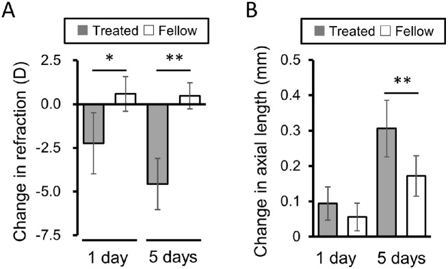
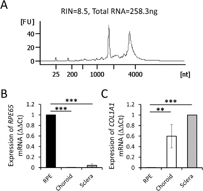
Methods: To induce myopia, two-week-old pigmented guinea pigs (New Zealand strain, n = 5) wore -10 diopter (D) rigid gas-permeable contact lenses (CLs), for one day; fellow eyes were left without CLs and served as controls. Spherical equivalent refractive errors (SE) and axial length (AL) were measured at baseline and one day after initiation of CL wear. RNA sequencing was applied to RPE collected from both treated and fellow (control) eyes after one day of CL-wear to identify related gene expression changes. Additional RPE-RNA samples from treated and fellow eyes were subjected to quantitative real-time PCR (qRT-PCR) analysis for validation purposes.

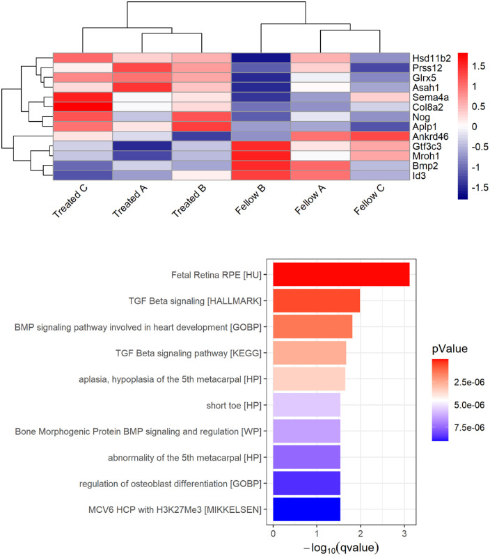
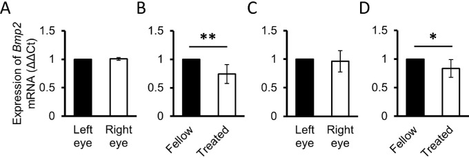
Results: The CLs induced myopia. The change from baseline values in SE was significantly different (P = 0.016), whereas there was no significant difference in the change in AL (P = 0.10). RNA sequencing revealed significant interocular differences in the expression in RPE of 13 genes: eight genes were significantly upregulated in treated eyes relative to their fellows, and five genes, including bone morphogenetic protein 2 (Bmp2), were significantly downregulated. The latter result was also confirmed by qRT-PCR. Additional analysis of differentially expressed genes revealed significant enrichment for bone morphogenetic protein (BMP) and TGF-β signaling pathways.

Conclusions: The results of this RPE gene expression study provide further supporting evidence for an important role of BMP2 in eye growth regulation, here from a guinea pig myopia model.


Conflict of interest statement

Disclosure: S. Goto, None; S.E. Muroy, None; Y. Zhang, None; K. Saijo, None; S.R.R. Kolora, None; Q. Zhu, None; C.F. Wildsoet, None

Figures

Figure 1.
Figure 1.
Changes from baseline values in refractive errors (A) and axial lengths (B) after one and five days of monocular imposed hyperopic defocus (−10 D contact lens), initiated at two weeks of age. * P < 0.05, ** P < 0.01.
Figure 2.
Figure 2.
Representative bioanalyzer output for RNA extracted from collected samples (A); and gene expression levels of RPE65, an RPE specific gene, and COL1A1, a choroid sclera-specific gene, measured by RT-qPCR in RPE, choroid and scleral samples from untreated animals (B and C, respectively), both data sets normalized to β-actin. ** P < 0.01; *** P < 0.001.
Figure 3.
Figure 3.
Effects of 1 day of exposure to hyperopic defocus (−10 D CL wear) on BMP and TGF-β signaling pathways in RPE; heatmap showing relative expression levels (z-scores) of genes found to be significantly upregulated (top 8 rows) and downregulated (bottom 5 rows) in RNA-seq analysis of treated versus fellow eyes. FDR < 0.1 (Top panel). Top 10 GSEA categories for the 13 differentially expressed genes include enriched biological pathways related to BMP and TGF-β signaling, and cell type enrichment for fetal RPE (Bottom panel). Categories are ranked by significance (decreasing −log10 [q value]) and color represents P values. The GSEA gene set for each category is listed in brackets. n = 3 for treated and fellow eyes (from 3 female animals), respectively. A, B, and C denote data from individual guinea pigs.
Figure 4.
Figure 4.
mRNA expression levels in RPE normalized to Gapdh (A and B) and β-actin (C and D) for Bmp2 from 15-day-old animals. (A) Bmp2 gene expression in RPE from untreated animals; there is no difference between right and left eyes (n = 3 animals) normalized to Gapdh. (B) Bmp2 gene expression after 1 day of monocular −10 D CL wear is significantly downregulated (n = 7 animals) normalized to Gapdh. (C) Bmp2 gene expression in RPE from untreated animals; there is no difference between right and left eyes (n = 3) normalized to β-actin. (D) Bmp2 gene expression after one day of monocular −10 D CL wear is significantly downregulated (n = 7 animals) normalized to β-actin. * P < 0.05, ** P < 0.01.

Similar articles

Cited by

References

    1. Holden BA, Fricke TR, Wilson DA, et al. .. Global prevalence of myopia and high myopia and temporal trends from 2000 through 2050. Ophthalmology. 2016; 123: 1036–1042. - PubMed
    1. Dolgin E. The myopia boom. Nature. 2015; 519(7543): 276–278. - PubMed
    1. Flitcroft DI. The complex interactions of retinal, optical and environmental factors in myopia aetiology. Prog Retin Eye Res. 2012; 31: 622–660. - PubMed
    1. Stone RA, Lin T, Laties AM, Iuvone PM.. Retinal dopamine and form-deprivation myopia. Proc Natl Acad Sci USA. 1989; 86: 704–706. - PMC - PubMed
    1. Seko Y, Shimizu M, Tokoro T.. Retinoic acid increases in the retina of the chick with form deprivation myopia. Ophthalmic Res. 1998; 30: 361–367. - PubMed

Publication types

Substances