. 2022 Jul 27;8(4):1947-1958.
doi: 10.3390/tomography8040163.

Partial Anomalous Left Pulmonary Artery Anterior Versus Posterior Types: A Systematic Review

Affiliations

Partial Anomalous Left Pulmonary Artery Anterior Versus Posterior Types: A Systematic Review

Carlos S Restrepo et al. Tomography. .

Abstract

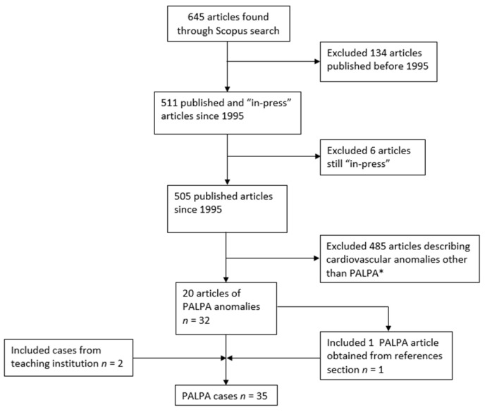
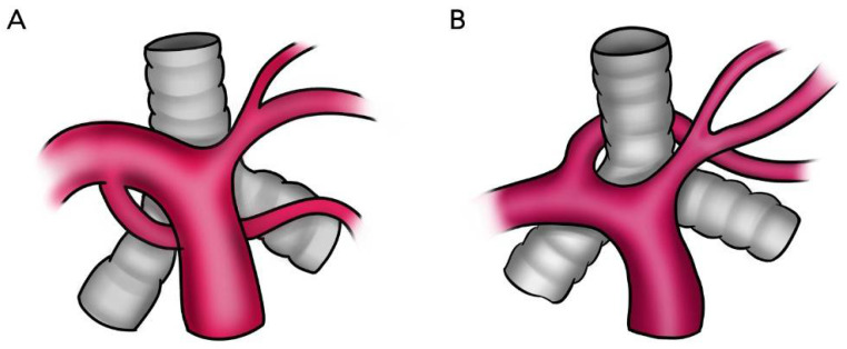
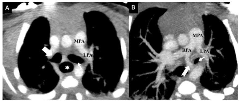
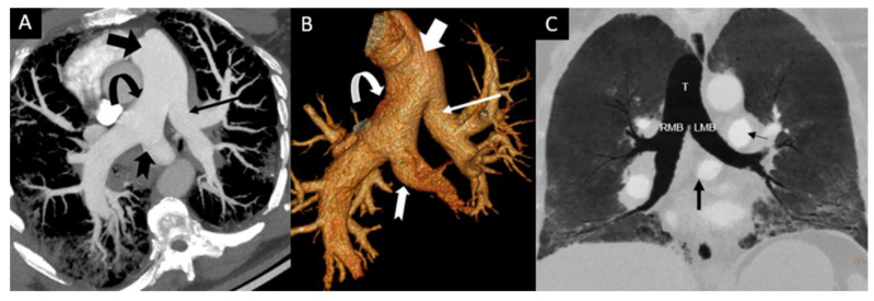
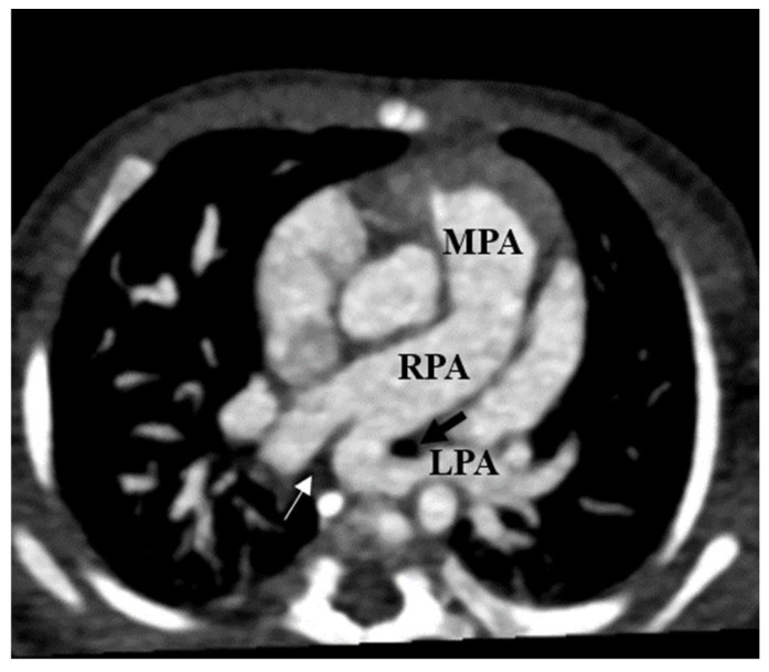
The aim of this study was to investigate the features of partial anomalous left pulmonary artery (PALPA) and differences between cases with posterior versus anterior a nomalous vessels in relation to the tracheobronchial tree. We hypothesized that statistical significance was dependent on the course of the anomalous vessel due to airway compression in the posterior type. This study included cases obtained from the literature (n = 33) and an institution teaching file (n = 2). Information collected: age, sex, medical history, additional anomalies, anomalous vessel course, and respiratory symptoms. Data were analyzed with independent samples t-test and Fisher's exact test. PALPAs were more commonly anterior than posterior. Mean age: 5.3 years (SD = 12.4) for anterior and 6.8 years (SD = 18.5) for posterior (p = 0.77). Respiratory symptoms: 20% of anterior and 60% of posterior cases (p = 0.032). Tracheobronchial anomalies: 35% of anterior and 60% of posterior cases (p = 0.182). Non-cardiac and non-tracheobronchial anomalies: 30% of anterior and 47% of posterior cases (p = 0.511). Kabuki syndrome: 25% of anterior and 6.7% of posterior cases (p = 0.207). In conclusion, respiratory symptoms were the only significant difference between anterior and posterior PALPA types.

Keywords: Kabuki syndrome; congenital heart disease; duplicated left pulmonary artery; partial anomalous left pulmonary artery (PALPA); pseudopulmonary artery sling.


Conflict of interest statement

The authors declare no conflict of interest.

Figures

Figure 1
Figure 1
Flowchart depicting systematic article exclusion and overall PALPA case acquisition from the literature and a single teaching institution. * = including but not limited to partial anomalous pulmonary venous connection, left pulmonary artery sling and anomalous coronary artery connection.
Figure 2
Figure 2
Schematic diagram of two representative patterns of PALPA branching and their anatomic relation with the tracheobronchial tree. (A) The anomalous duplicated left pulmonary artery originates from the inferior aspect of the right pulmonary artery and passes to the left anteriorly to the tracheobronchial tree. (B) The anomalous duplicated left pulmonary artery arises from the posterior–superior right pulmonary artery and passes to the left hilum between the trachea and the esophagus (not shown).
Figure 3
Figure 3
Newborn male with complex congenital heart disease (double outlet right ventricle, ventricular septal defect, and aortic coarctation) with PALPA. Contrast enhanced axial (A) and (B) axial oblique reconstructed Maximum Intensity Projection (MIP) images from chest CT data demonstrate a duplicated left pulmonary artery with a normally arising left upper lobe pulmonary artery (LPA) from the main pulmonary artery (MPA) coursing anterior to the left main bronchus (thin small arrow), while an anomalous left lower lobe pulmonary artery (notched arrow) coursing inferior anterior to the left main bronchus and left lateral to the esophagus (star). No anomalous vessel was seen between the trachea and the esophagus. Right main bronchus is marked with a bold white arrow.
Figure 4
Figure 4
Incidental finding of PALPA in a 51-year-old male with acute shortness of breath. Contrast enhanced chest CT obtained to rule out pulmonary embolism demonstrates that a duplicated left pulmonary artery with a small left pulmonary artery (thin arrow) to left upper lobe was arising from the main pulmonary artery (bold arrow) and an additional anomalous left pulmonary artery to the left lower lobe (notched arrow) was arising from the proximal posterior part of the right pulmonary artery (curved arrow) on axial oblique reconstructed Maximum Intensity Projection (MIP) image (A) and Volume Rendered reconstructed image (B). There was no associated congenital heart disease or tracheobronchial stenosis. Coronal Minimum intensity projection (MinIP) (C) reconstructed image from same patient shows that the course of anomalous LPA branch (PALPA, bold black arrow) is caudal to the carina and is inferior anterior and parallel to the left main bronchus (LMB). There was no tracheal (T), right main (RMB) or left main bronchial (LMB), or esophageal compression. Note that left upper lobe and the lingula were supplied by the proximal left pulmonary artery (thin black arrow) which arises in the usual position from the main pulmonary artery (MPA). Note peripheral reticular opacities and ground glass opacities in both lungs related to known non-specific interstitial pneumonia (NSIP).
Figure 5
Figure 5
Left pulmonary artery sling (LPAS) in a 3-month-old girl with a cardiac murmur. Axial thin chest CTA shows an anomalous left pulmonary artery (LPA) arising from the posterior part of the proximal right pulmonary artery (RPA) and coursing between the left main bronchus (black arrow) and esophagus (thin white arrow) to the left pulmonary hilum.

Similar articles

Cited by

References

    1. Maldjian P.D., Adams K.R. Partial anomalous left pulmonary artery sling in an adult. J. Clin. Imaging Sci. 2020;10:5. doi: 10.25259/jcis_4_2020. - DOI - PMC - PubMed
    1. Sen S., Winlaw D.S., Sholler G.F. Partial anomalous left pulmonary artery: Report of two cases and review of literature. Cardiol. Young. 2015;25:1012–1014. doi: 10.1017/S1047951114001528. - DOI - PubMed
    1. Wang S.Y., Gao W., Zhong Y.M., Sun A.M., Wang Q., Hu L.W., Qiu H.S., Li J.Y., Jaffe R.B. Multislice computed tomography assessment of tracheobronchial patterns in partial anomalous left pulmonary artery. J. Comput. Assist. Tomogr. 2017;41:983–989. doi: 10.1097/RCT.0000000000000623. - DOI - PubMed
    1. Xie J., Juan Y.H., Wang Q., Chen J., Zhuang J., Xie Z., Liang C., Zhu Y., Yu Z., Li J., et al. Evaluation of left pulmonary artery sling, associated cardiovascular anomalies, and surgical outcomes using cardiovascular computed tomography angiography. Sci. Rep. 2017;7:40042. doi: 10.1038/srep40042. - DOI - PMC - PubMed
    1. Yuan S.M. Congenital heart defects in Kabuki syndrome. Cardiol. J. 2013;20:121–124. doi: 10.5603/CJ.2013.0023. - DOI - PubMed

Publication types